
In the delicate, labyrinthine landscape of the human brain, the ability to observe, measure, and influence neural activity is the holy grail of neuroscience. For decades, researchers have been hampered by the limitations of "blunt" tools—rigid, silicon-based probes that offer a limited view of the brain’s complex, multi-layered architecture. Now, a groundbreaking development from a multidisciplinary team of scientists at DTU (Technical University of Denmark), the University of Copenhagen, and University College London is poised to shift the paradigm.
The team has unveiled the "microfluidic Axialtrode" (mAxialtrode), a needle-thin, flexible brain implant that promises to revolutionize how we study neurological disorders like epilepsy and memory loss. By integrating fluid delivery, electrical recording, and optical stimulation into a single, hair-thin fiber, the mAxialtrode represents a quantum leap in neuro-engineering.
Main Facts: A Convergence of Technologies
The mAxialtrode is not merely an electrode; it is a comprehensive, multi-modal interface between technology and biological tissue. Published in the journal Advanced Science, the study describes a device that fundamentally alters the spatial resolution of brain research.
The implant is constructed from soft, biocompatible polymers, allowing it to mimic the mechanical properties of brain tissue. Unlike traditional rigid silicon probes—which are prone to causing chronic inflammation and "gliosis" (scarring) due to their hardness—the mAxialtrode moves in tandem with the brain. Its architecture is a marvel of miniaturization: a central light-conducting core is ringed by eight microscopic fluidic channels, which can also house ultra-thin metal wires for recording electrical signals.
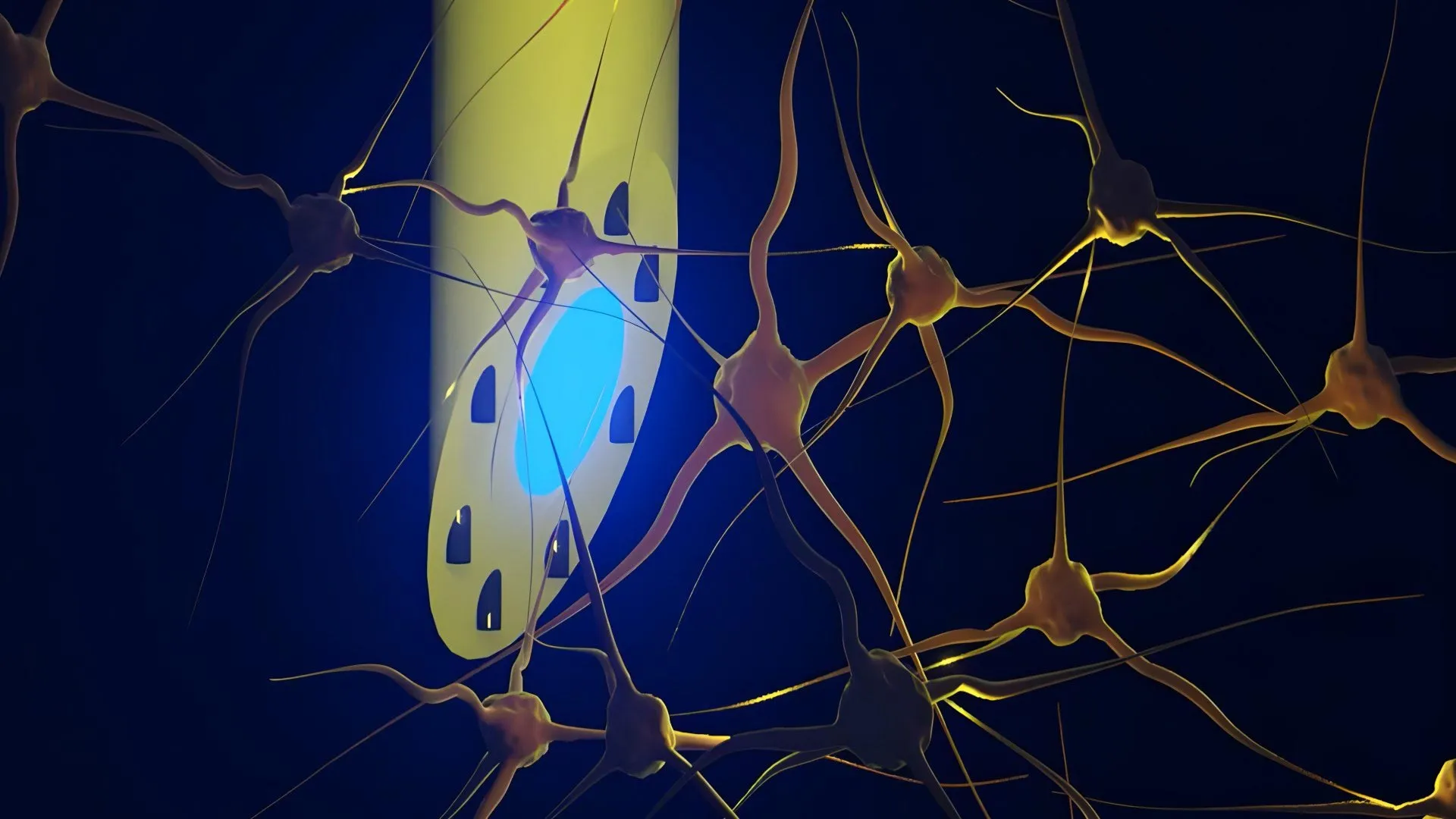
The device is less than half a millimeter in diameter, yet it offers functionality across its entire length. This allows researchers to stimulate neurons with light (optogenetics), record electrical spikes (electrophysiology), and deliver precise chemical payloads (pharmacology) at varying depths, all from a single insertion point.
Chronology: From Polymer Rod to Living Tissue
The journey to the mAxialtrode began with a fundamental question: how do we increase the "functional density" of a brain implant without increasing its physical size?
- Conceptualization (The Development Phase): Postdoc Kunyang Sui and Associate Professor Christos Markos began by challenging the standard manufacturing processes for optical fibers. They utilized a "thermal drawing" process—a technique akin to stretching molten glass into a fine strand, but applied here to high-precision polymers.
- The Prototyping Phase: The researchers experimented with various configurations, ultimately settling on a geometry that could incorporate multiple micro-channels without compromising the flexibility or the optical integrity of the fiber.
- Initial Laboratory Validation: Before moving to living systems, the team conducted rigorous benchtop testing to ensure the integrity of the fluidic channels and the conductivity of the electrical wires.
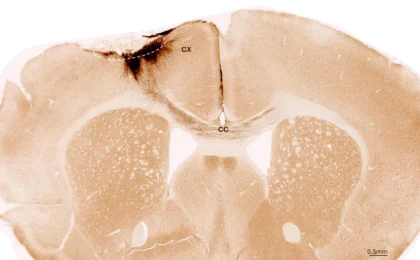
- In Vivo Testing: Recognizing that a device is only as good as its performance in a living system, the team collaborated with neurophysiologists Associate Professor Rune W. Berg and Associate Professor Rob C. Wykes. The team performed in vivo tests in mice. They successfully demonstrated that the implant could deliver blue and red light for optogenetics, record signals from both the cerebral cortex and the hippocampus, and inject substances at specific, distinct depths up to three millimeters apart.
Supporting Data: Why Current Methods Fail
To understand the magnitude of the mAxialtrode’s impact, one must look at the limitations of current research tools. For years, the gold standard in optogenetics has been the flat-ended optical fiber. These fibers are highly effective at delivering light to a single, localized point, but they are essentially "blind" to the layers surrounding that point.
The "Nose" Limitation
In traditional fiber-optic implants, light is emitted only from the "distal tip" or the nose of the fiber. This creates a binary, single-point interaction. If a researcher wants to study how a signal travels from the outer layers of the cerebral cortex down to the deep-seated hippocampus, they would typically need to insert multiple, bulky probes. This not only increases the risk of damage to the brain but also introduces "noise" into the data, as multiple devices can distort the natural neural architecture they are trying to monitor.
Mechanical Mismatch
The "stiffness mismatch" between silicon-based electrodes and the soft, jelly-like consistency of the brain is a significant hurdle in long-term implantation. Rigid materials cause the brain to recoil during movement, leading to constant micro-trauma. The mAxialtrode’s soft polymer design mitigates this, promising a future where implants can reside in the brain for months or even years without triggering the body’s natural inflammatory defense system.
Official Responses and Expert Perspective
The research team, while optimistic, maintains a professional, cautious stance regarding the transition from the laboratory to the hospital ward.
"Most current brain implants are based on hard materials such as silicon, which can irritate the brain and trigger inflammatory reactions in the tissue," says Kunyang Sui. "The new implant differs in that it is made of soft, plastic-like optical fibers and has a specially angled tip that makes it smaller and reduces the damage caused when it is placed in the brain."
Associate Professor Christos Markos emphasizes the utility of the "all-in-one" approach. "By combining several capabilities—electrical, optical, and chemical—into a single implant, we reduce the footprint of our interventions. This is not just about convenience; it is about data fidelity. When you insert fewer probes, you get a clearer, more accurate picture of the brain’s natural state."
However, the researchers are quick to temper expectations. "This is a profound advancement for neuroscience research," Sui adds, "but we are still far from routine clinical use. We need extensive long-term safety testing, further refinement of the manufacturing process, and rigorous regulatory approval before this could ever be considered for a human patient."
Implications: A New Era for Epilepsy and Neurology
The potential therapeutic applications of the mAxialtrode are vast, particularly for conditions that are currently difficult to manage with systemic medication.
Targeted Epilepsy Treatment
Epilepsy is often characterized by the misfiring of neural circuits in specific brain regions. Currently, treatment often involves systemic anti-epileptic drugs, which circulate throughout the entire body, causing a host of side effects. The mAxialtrode could enable "closed-loop" systems. In such a scenario, the device would detect the electrical signature of an impending seizure and immediately deliver a precise dose of medication directly to the hyper-excitable tissue, effectively "quenching" the seizure before it manifests.
Memory and Decision-Making Research
Beyond treatment, the device provides an unprecedented window into the fundamental mysteries of the human mind. By allowing scientists to manipulate and monitor neural activity across multiple layers of the cortex and deep structures simultaneously, the mAxialtrode could help map the neural circuits responsible for memory formation, decision-making, and emotional regulation.
Next Steps: Toward Clinical Translation
The team is currently working to patent the technology, an essential step in securing the investment and resources required for clinical development. Their roadmap includes:
- Miniaturization refinement: Ensuring the manufacturing process is scalable and consistent.
- Long-term Biocompatibility Studies: Testing the device over longer durations to ensure no degradation or chronic inflammation occurs.
- Human-relevant Models: Partnering with clinical neurologists to identify the specific patient populations that would benefit most from this localized, multi-modal approach.
The mAxialtrode is a testament to the power of interdisciplinary science. By bridging the gap between polymer physics and neurophysiology, researchers have created a tool that respects the complexity of the brain rather than fighting against it. As the project moves toward the patenting and clinical exploration phases, the scientific community watches with anticipation, hopeful that this needle-thin thread may one day weave a new tapestry of treatment for the most complex organ in the human body.