

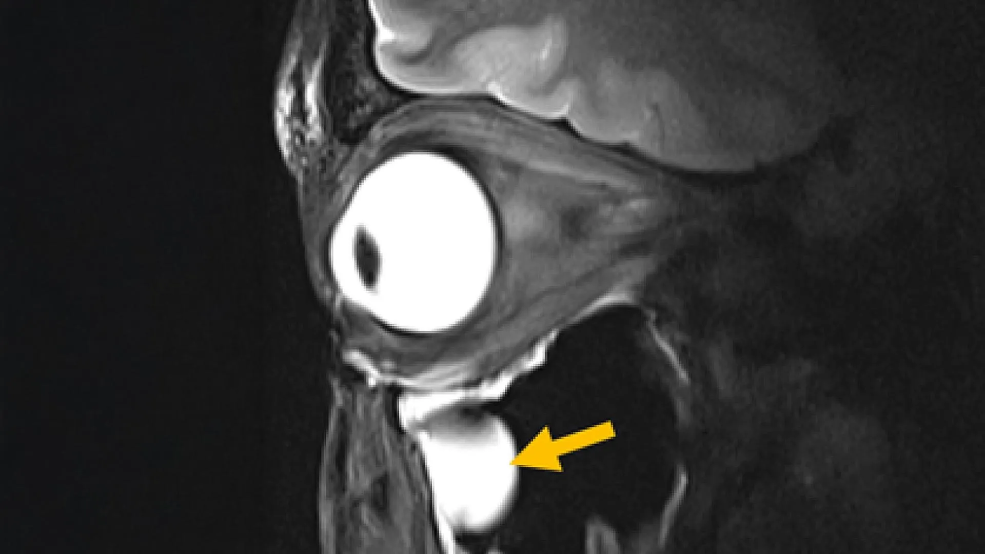
Magnetic resonance imaging (MRI) has long been the gold standard for non-invasive medical diagnostics, providing clinicians with a high-definition window into the human body’s internal landscape. Yet, despite decades of iterative refinement, the technology has hit a persistent ceiling: the hardware responsible for transmitting and receiving radiofrequency (RF) signals—the "eyes and ears" of the MRI machine—often struggles to capture high-quality data from deep-seated brain structures or delicate, small-scale regions like the human eye and its surrounding orbital cavity.
Today, a groundbreaking development led by Nandita Saha, a doctoral student in Professor Thoralf Niendorf’s Experimental Ultrahigh Field Magnetic Resonance laboratory at the Max Delbrück Center, promises to shatter those limitations. By integrating advanced metamaterials into traditional MRI antenna designs, the research team has successfully demonstrated a method to produce sharper, faster, and more detailed images without the need for prohibitively expensive infrastructure upgrades. Published in the journal Advanced Materials, this breakthrough marks a significant leap toward a new era of diagnostic precision.
The Technical Hurdle: Why MRI Struggles with Complex Anatomy
To understand the magnitude of this innovation, one must first grasp the mechanics of an MRI scan. An MRI scanner operates by bathing the body in powerful magnetic fields and pulsing radiofrequency signals into the patient’s tissues. As atoms in the body—typically hydrogen protons—resonate with these signals, they emit an electromagnetic response. The scanner’s RF coils (antennas) detect these echoes and translate them into the detailed images clinicians use to diagnose tumors, neurological disorders, and vascular issues.
The primary challenge lies in the "signal-to-noise ratio" (SNR). In anatomically complex areas like the eye, the signal is often faint. Traditional RF coils are bulky and struggle to focus electromagnetic energy precisely. When the signal is weak, the resulting image becomes "noisy" or blurred, often necessitating longer scan times to acquire enough data for a clear picture. For patients, this translates to longer periods of immobility inside a loud, confined machine—an experience that is both physically uncomfortable and prone to motion-induced errors.
A New Frontier: The Metamaterial Solution
The research team, a collaborative effort involving experts in MRI physics, clinical ophthalmology, and translational imaging from the Max Delbrück Center and Rostock University Medical Center, turned to the burgeoning field of metamaterials to solve the SNR problem.
Metamaterials are synthetic structures engineered to exhibit properties not found in naturally occurring materials. By manipulating the geometric configuration of these materials at a scale smaller than the wavelength of the RF signals, the researchers can control how electromagnetic waves propagate.
"By using concepts from metamaterials, we were able to guide radiofrequency fields more efficiently and demonstrate how advanced physics can directly improve medical imaging," explains Professor Thoralf Niendorf, the study’s senior author. "This work shows a pathway toward faster, clearer MRI scans that could benefit patients in many clinical areas."
In practical testing, the researchers incorporated these metamaterial structures directly into the MRI antenna. When applied to 7.0 Tesla (T) MRI systems, the antenna acted as a signal "lens," focusing RF energy into the targeted tissues. This resulted in a marked increase in spatial resolution and significantly accelerated data collection, effectively solving the "blur" issue that has historically plagued ophthalmological and deep-brain imaging.
Chronology of the Research and Validation
The project was not a solitary endeavor but a multi-year, multi-institutional initiative designed to bridge the gap between theoretical physics and clinical reality.
- Initial Conceptualization: The team, led by Saha and Niendorf, began by reimagining the antenna from the perspective of modern electromagnetic theory, aiming to move beyond traditional copper-based coil designs.
- Prototyping: Working in the Max Delbrück Center’s ultrahigh field laboratory, the team designed compact, lightweight metamaterial-integrated antennas.
- Validation Phase: The team partnered with University Medicine Rostock to validate the design in a clinical environment. Professor Oliver Stachs and Dr. Ebba Beller provided the clinical oversight necessary to ensure the technology could function effectively on living human subjects.
- Clinical Testing: The team successfully utilized the new antenna to image the eyes and orbits of human volunteers at 7.0 Tesla. The results confirmed that the technology was not only functional but superior to conventional equipment.
- Publication and Peer Review: The findings were subjected to rigorous scrutiny and subsequently accepted by Advanced Materials, validating the methodology and the potential impact of the technology.
Official Perspectives: The Clinical Impact
The collaboration between the Max Delbrück Center and the University Medicine Rostock emphasizes the importance of translational medicine—taking laboratory physics and applying them directly to patient care.
Professor Oliver Stachs, a co-author of the study, notes that the impact on ophthalmology is potentially transformative. "Our research demonstrates clear relevance for ophthalmological applications as it can facilitate anatomically detailed, high-spatial resolution MRI of the eye," says Stachs. "It offers the potential to open a window into the eye and into (patho)physiological processes that in the past have been largely inaccessible."
For clinicians, this means being able to visualize minute structural changes in the eye that were previously obscured by image artifacts. This could lead to earlier detection of diseases such as ocular tumors, glaucoma, or inflammatory conditions that require precise, non-invasive observation.
Dr. Ebba Beller, also of the Rostock University Medical Center, views this study as a foundational step. "Innovations in imaging hardware have the potential to transform diagnostics, and this study is an important step toward next-generation MRI technology," she says.
Implications: Beyond the Eye and Orbit
While the current study focuses on the eye and orbit, the researchers emphasize that the underlying physics are highly adaptable. The goal, as Nandita Saha puts it, was to "rethink MRI hardware from the modern physics of antenna design."
Enhanced Patient Comfort and Efficiency
Because the new antenna is lightweight and compact, it can be tailored to the specific geometry of different body parts. This allows for more localized, ergonomic designs that increase patient comfort. Furthermore, the efficiency of the signal capture means shorter scan sessions. Shorter scans reduce the likelihood of patient movement, which in turn reduces the need for repeat scans, lowering costs for healthcare providers and reducing stress for patients.
Safety and Specialized Imaging
The metamaterial approach offers unique safety benefits. By guiding RF energy more precisely, the antenna can potentially reduce "hot spots"—unintended areas of heating caused by electromagnetic energy. This is a critical safety feature, particularly for patients with metallic medical implants who might otherwise be barred from high-field MRI scans.
Additionally, the technology holds promise for "non-proton" MRI. While most scans focus on hydrogen atoms, researchers are increasingly interested in tracking sodium or fluorine, which can provide deeper insights into metabolic processes. The improved sensitivity provided by the metamaterial antenna could make these specialized techniques more viable and routine.
Future Trajectories: Scaling and Adaptation
The research team is not resting on these initial successes. They have outlined a clear roadmap for the future of this technology, which includes:
- Expanded Clinical Studies: The team is currently preparing for larger, multi-hospital studies to test the antenna’s efficacy across a broader range of patient demographics.
- Organ-Specific Adaptation: Development is underway to adapt the metamaterial structures for imaging other complex organs, specifically the heart and the kidneys, where precise imaging is vital for monitoring chronic conditions.
- Cross-Field Compatibility: Professor Niendorf notes that the design is modular. It can eventually be adapted for use in lower-field (e.g., 1.5 T or 3.0 T) and even higher-field MRI systems, potentially democratizing access to high-quality diagnostic imaging.
- Reciprocal Collaboration: The partnership between the Max Delbrück Center and Rostock University Medical Center will continue through ongoing scientist exchange programs, ensuring that the development cycle remains tightly linked to clinical needs.
Conclusion: A New Standard for Diagnostics
The integration of metamaterials into MRI hardware represents more than just an incremental update to existing machinery. It is a fundamental shift in how we approach electromagnetic wave management in medicine. By moving away from the "one-size-fits-all" coil design and toward precision-engineered antennas, researchers are unlocking higher resolutions and faster speeds.
As this technology matures and moves from the laboratory to the hospital, it promises to sharpen our view of the human body, turning once-blurry diagnostic images into high-definition maps of disease. With the backing of the DFG (German Research Foundation) and the dedication of researchers like Saha, Niendorf, and Stachs, the future of MRI is not just faster or more comfortable—it is fundamentally more capable of revealing the invisible.