

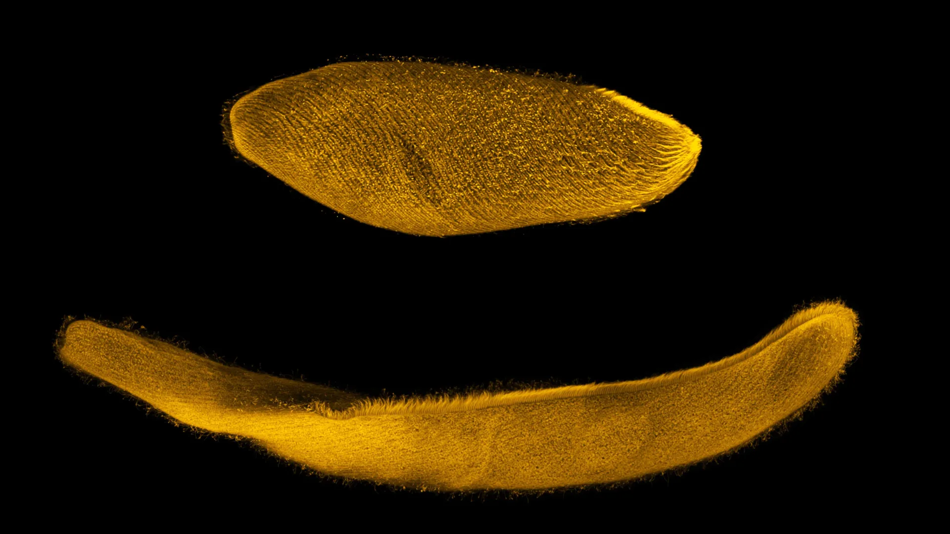
In the quiet, microscopic realm of freshwater ponds and stagnant pools, a biological marvel performs a feat of physics that defies conventional understanding of cellular movement. Spirostomum ambiguum, a giant single-celled ciliate, possesses the extraordinary ability to compress its elongated body to one-quarter of its original length in under five milliseconds. To put this into perspective, the organism performs this rapid contraction hundreds of times faster than a human can blink. This singular event, occurring at a speed of roughly 100 body lengths per second, has long captivated biologists. Now, an interdisciplinary team of researchers has finally decoded the mechanical "fishnet" that powers this high-speed performance, potentially paving the way for a new generation of synthetic, high-performance artificial muscles.
The Mechanics of a Microscopic Athlete
At first glance, Spirostomum appears as a simple, worm-like organism covered in a fringe of cilia—the hairlike organelles that propel it through the water. However, beneath its translucent membrane lies a sophisticated kinetic architecture that differentiates it from almost every other known single-celled entity. While human muscle fibers rely on a complex chemical cascade to shorten, Spirostomum operates with a level of efficiency and speed that human biology simply cannot match.
The core of this research, published in the Proceedings of the National Academy of Sciences, highlights the organism’s reliance on "myonemes"—fibrous structures that function as a biological contractile system. Unlike the sarcomeres found in human muscle tissue, which are powered by the energy-dense molecule adenosine triphosphate (ATP), Spirostomum utilizes a calcium-ion-triggered mechanism. This process is essentially "electric" in nature, operating with a speed and repeatability that suggests an entirely different evolutionary trajectory for movement.
A Chronology of Discovery: From Observation to Analysis
The study of Spirostomum has been a multi-year endeavor involving experts in biophysics, chemistry, and biological engineering. The research team, led by Mary Elting of North Carolina State University, along with collaborators from the University of Chicago, Drake University, and the University of Colorado at Boulder, approached the problem by integrating high-resolution microscopy with theoretical modeling.
The Initial Inquiry
The project began with a fundamental question: How does a single cell, lacking a nervous system or traditional muscle fibers, execute a movement so violent and rapid that it would tear most cells apart? Initial observations confirmed that the contraction was not merely a reaction to external stimuli, but a highly orchestrated physical event.
Mapping the Fishnet
Using a combination of electron microscopy and immunofluorescence, the researchers visualized the interior of the organism. They discovered a network of proteins—specifically centrin and Sfi1—arranged in a geometric pattern that resembled a fishnet. This network encases the cell, providing both the structural integrity required to survive the force of the contraction and the flexibility to initiate it.
Identifying the Trigger
By manipulating the internal environment of the cell, the team confirmed that calcium ions act as the primary catalyst. When these ions are introduced, the Sfi1 protein undergoes a conformational shift, changing from a rigid, structural component into a pliable, clumped mass. This "wet spaghetti" transformation forces the fishnet to pull inward, causing the organism to collapse in on itself in a fraction of a second.
Supporting Data: The Physics of Contraction
To understand why this organism is so unique, one must compare it to the standard model of animal movement. Human muscles are powered by ATP, which is "burned" to facilitate the sliding of actin and myosin filaments. This is a robust but relatively slow chemical process.
The researchers characterize the difference between human muscle and Spirostomum as analogous to the difference between internal combustion and electric power. In the organism, the contraction is triggered by a sudden influx of calcium, which creates an electrical-like shift in the Sfi1 proteins. Because the fishnet geometry allows for a uniform, distributed contraction, the organism’s delicate internal organelles—the equivalent of organs—are shielded from the immense forces generated during the rapid shortening of the body.
The data gathered by the research team showed that this process is not just a "one-shot" mechanism. Unlike many other explosive biological movements, such as the firing of a nematocyst in jellyfish or the snap of a Venus flytrap, Spirostomum is capable of repeating this motion almost instantaneously. This suggests that the cell possesses a rapid "reset" mechanism, allowing it to recharge its calcium stores or reconfigure its Sfi1 proteins almost as quickly as it fires.
Official Responses and Expert Insights
Mary Elting, associate professor of biophysics at NC State and co-corresponding author of the study, emphasizes that the discovery is more than a biological curiosity—it is a blueprint for future technology.
"The difference between what Spirostomum can do and what we can do comes down to what is powering the contraction, and what the machinery behind it looks like," Elting explains. "If we can understand those processes, it could help us build synthetic systems that mimic the speed and power of this single-celled organism."
The collaboration highlights the necessity of interdisciplinary work. By bringing together experts like Aaron Dinner (University of Chicago), Jerry Honts (Drake University), and Saad Bhamla (University of Colorado at Boulder), the team was able to bridge the gap between microscopic observation and macro-scale engineering.
"We would expect calcium-triggered reactions to be ‘one shot,’ but Spirostomum can do it repeatedly," Elting notes. "Understanding those aspects of its motion are the keys to building a fast-moving, ATP-independent artificial muscle."
Implications for Synthetic Biology and Engineering
The potential applications for this research are profound. Current artificial muscle technologies—often based on soft robotics or pneumatic actuators—are frequently limited by weight, power source requirements, and speed. By mimicking the calcium-driven, fishnet-structured myonemes of Spirostomum, engineers may be able to develop:
- High-Speed Synthetic Actuators: Devices that can move with the speed of an insect but the durability of an industrial component.
- ATP-Independent Robotics: By shifting from chemical combustion (ATP) to electrical-ion signaling, robots could potentially operate in environments where traditional batteries are impractical or where rapid bursts of energy are required.
- Micro-scale Medical Devices: The fishnet geometry could inspire new ways to protect sensitive payloads (such as drug delivery systems) inside synthetic capsules that need to deform to navigate the human vascular system.
Unanswered Questions and Future Directions
Despite the breakthrough in identifying the fishnet structure and the Sfi1 protein’s role, significant mysteries remain. The research team is now turning its attention to the "reset" mechanism. How does the organism clear the calcium ions to return to its original state so quickly? What generates the voltage that initiates the current in the first place?
The answers to these questions will likely involve further study into the cell’s membrane potential and the ion channels that govern the flow of calcium. As the team continues to refine their understanding, they hope to transition from biological observation to synthetic implementation.
The research represents a significant leap in our understanding of cellular biomechanics. It serves as a reminder that the natural world, even at the scale of a single cell, continues to hold lessons for engineers and scientists alike. By decoding the "fishnet" of Spirostomum ambiguum, humanity has taken a step closer to creating machines that move with the same fluid, explosive, and efficient power as the smallest athletes on Earth.
Acknowledgments and Funding
The research, titled “The fishnet geometry of the myoneme in Spirostomum ambiguum enables rapid, uniform, and repeatable contraction,” received broad support from the scientific community. Funding was provided by the National Science Foundation (Award numbers 1935260, 2313722, 2313724, 1935262, 1817334, 2313727, and 2313725) and the National Institutes of Health (Award numbers R35GM130327 and R35GM142588).
The research team included Joseph Lannan and Peter Thompson of NC State; Carlos Floyd and Suriyanarayanan Vaikuntanathan of the University of Chicago; L.X. Xu of the Georgia Institute of Technology; and Connie Yan and Wallace Marshall of the University of California San Francisco. Through this collaborative effort, the team has provided a new lens through which we view the limits of speed and the potential of biological machinery.