
In a landmark development for oncology, researchers at Weill Cornell Medicine and the Cornell Duffield College of Engineering have unveiled a novel therapeutic approach that utilizes "ultrasmall" silica nanoparticles to eradicate aggressive prostate tumors. Published in the June 15 issue of the journal Cancer Research, the study reveals that these engineered particles, known as Cornell Prime dots (C’ dots), possess the dual capability to directly trigger tumor cell death while simultaneously priming the immune system to recognize and attack the malignancy.
This preclinical study, which demonstrated complete tumor remissions in mouse models, represents a potential paradigm shift in cancer care. By operating through multiple biological pathways at once, these nanoparticles may overcome the historical resistance that has long plagued prostate cancer treatments, offering a promising roadmap toward human clinical trials.
The Science of the "C’ Dot": A Multi-Pronged Attack
At the heart of this innovation are amorphous silica nanoparticles—a form of silicon dioxide that occurs naturally in many foods and the fossilized remains of microscopic organisms. While these particles were originally engineered by the laboratory of Dr. Ulrich Wiesner for medical imaging, their potential as a therapeutic agent has emerged through an interdisciplinary collaboration with the laboratory of Dr. Michelle Bradbury.
Unlike traditional chemotherapy, which often relies on systemic toxicity to kill cancer cells, C’ dots employ a sophisticated "Trojan horse" strategy. The particles are designed to carry a targeting molecule that recognizes PSMA (Prostate-Specific Membrane Antigen), a protein highly expressed on the surface of prostate tumor cells. This ensures that the therapy accumulates precisely where it is needed most, minimizing exposure to healthy tissue.
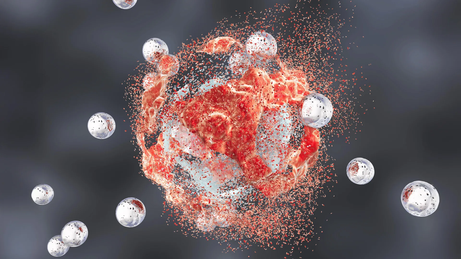
Once internalized by the tumor cell, the nanoparticles initiate a process known as "ferroptosis"—a specialized form of cell death driven by overwhelming oxidation. Evidence suggests that the nanoparticles act as a transport vehicle, gathering positively charged iron ions from the bloodstream and ferrying them into the tumor cell. Once inside, these iron ions catalyze intense oxidative stress, damaging the lipid-rich membranes of the cancer cells until they collapse and disintegrate.
Chronology of Discovery: From Imaging to Immunotherapy
The journey of the C’ dot began over a decade ago as a tool for precision medical imaging. Because of their ultrasmall size and unique chemical structure, researchers found that these particles could navigate the body’s circulatory system and highlight tumors during surgical procedures with unparalleled clarity.
As these particles moved into late-stage clinical trials for image-guided surgery, the research team began to observe unexpected biological interactions. They noticed that the particles were not merely passive observers; they were actively interfering with the metabolic and survival pathways of cancer cells.
- Phase I: Discovery of Imaging Potential. Researchers developed C’ dots as highly efficient, biocompatible markers for tumor visualization.
- Phase II: Identification of Biological Interaction. Observations during imaging studies suggested that the nanoparticles could selectively damage tumor cells without harming surrounding healthy tissues.
- Phase III: Mechanism Mapping. The team identified the ferroptosis pathway and the role of iron ions in fueling oxidative tumor death.
- Phase IV: Immune System Activation. The team discovered that the destruction of tumor cells released antigens that "woke up" dormant immune cells, shifting the tumor microenvironment from a "cold" (immune-resistant) state to a "hot" (immune-active) state.
- Phase V: Preclinical Validation. In 2024, the team successfully demonstrated that combining these particles with standard immunotherapy led to significant and, in some cases, complete tumor remission in aggressive prostate cancer models.
Supporting Data: Survival and Synergy
The most compelling evidence from the study lies in the survival outcomes observed in mice. Prostate cancer is notoriously difficult to treat with immunotherapy alone because the tumor microenvironment is often immunosuppressive, effectively hiding the cancer from the body’s T cells.
In the study, the researchers tested several treatment configurations:
- Monotherapy (C’ dots or immunotherapy alone): Both methods provided only modest improvements in survival rates compared to untreated control groups.
- Dual Therapy (C’ dots + Immune Checkpoint Blockade): The combination produced a synergistic effect, resulting in complete or nearly complete remissions and indefinite survival in 40% of the mice.
- Triple Therapy (C’ dots + Immune Checkpoint Blockade + CSF-1R Blockade): By adding a third agent to target tumor-associated macrophages, the researchers achieved complete remission in 50% of the subjects.
These findings are statistically significant, suggesting that the nanoparticles do more than just kill cells; they fundamentally reorganize the tumor environment to make it vulnerable to other therapeutic agents. By disrupting the metabolic processes that allow tumors to hide from the immune system, C’ dots effectively "unlock" the full potential of existing cancer drugs.
Official Responses and Expert Perspectives
The project is the result of a long-running partnership between Dr. Michelle Bradbury and Dr. Ulrich Wiesner, bringing together the worlds of radiology, oncology, and materials science.
"It seems unreal—how is it possible that rather than a single pathway, we see all these effects happening simultaneously and only in tumors and not in healthy tissues?" said Dr. Wiesner, the Spencer T. Olin Professor in the Department of Materials Science and Engineering at Cornell. "I have to wonder whether ultrasmall silica’s very early and ubiquitous presence in the environment and foods has given it a connection to biology that we’re only beginning to glimpse."
Dr. Bradbury, who serves as the director of the Molecular Imaging Innovations Institute at Weill Cornell Medicine, emphasized the clinical significance of the findings. "We’re very encouraged by these results; a treatment that directly induces tumor-cell death while transforming the immune microenvironment would represent a new clinical paradigm," she stated.
Dr. Jedd Wolchok, a co-author of the study and a leader at the Parker Institute for Cancer Immunotherapy, highlighted the broader implications for the field. "One of the most intriguing aspects of this work is the convergence of direct tumor cell killing with broad immune remodeling," Dr. Wolchok noted. "By creating conditions that support a more effective antitumor immune response, these particles may help unlock the full potential of immunotherapy in prostate cancer, where durable responses have historically been difficult to achieve."
Future Implications and Clinical Outlook
The transition from mouse models to human clinical trials remains the critical next step. The research team is currently focused on evaluating the safety profile of the nanoparticles in larger, more complex biological systems. While the accumulation of particles in the spleen was noted in the study, researchers emphasized that there were no signs of systemic toxicity, a common hurdle for nanoparticle-based therapies.
If successful in human trials, the C’ dot approach could provide a versatile platform for treating various types of solid tumors. Because the targeting mechanism is modular—meaning the "targeting molecule" can be swapped out to recognize different cancer types—the fundamental technology could be adapted for lung, breast, or pancreatic cancers.
The research also opens new doors in the study of ferroptosis. By learning how to modulate this form of cell death with surgical precision, scientists may be able to develop a new class of metabolic cancer therapies that are both potent and highly specific.
As the team prepares for the next phases of development, the collaboration between Weill Cornell Medicine and Cornell University serves as a testament to the power of interdisciplinary research. With the backing of the National Cancer Institute and the Department of Defense, the team is now moving toward translating these findings into a clinical reality that could fundamentally alter the landscape of prostate cancer treatment.
For patients who have exhausted traditional lines of care, the prospect of a therapy that turns the body’s own immune system into an active weapon against cancer offers a beacon of hope, grounded in the sophisticated application of materials science.