
In the dense, crowded environment of the living cell, the fundamental unit of heredity—DNA—faces a significant physical hurdle. Because the backbone of the DNA double helix is inherently negatively charged, the laws of physics dictate that these molecules should naturally repel one another, much like the opposing poles of a magnet. Yet, life relies on the ability of DNA strands to find, recognize, and interact with matching sequences. This "molecular recognition" is the cornerstone of genetic recombination, gene silencing, and DNA repair.
For decades, the mechanism behind this close-range attraction remained a theoretical mystery. Now, an international team of researchers has bridged the gap between theory and observation. By combining cutting-edge atomic force microscopy with high-resolution computer simulations, scientists have confirmed that positively charged metal ions act as "molecular bridges," allowing DNA strands to lock together with extraordinary precision. This discovery, published in the journal Nucleic Acids Research, validates a two-decade-old hypothesis and opens new doors for understanding cancer development and the future of synthetic biotechnology.
The Physics of Repulsion: A Decades-Old Enigma
To understand the significance of this breakthrough, one must first appreciate the scale of the challenge. DNA is a polyelectrolyte; its phosphate backbone is laden with negative charges. In an aqueous cellular environment, these charges create a powerful electrostatic repulsive force. Despite this, DNA must frequently navigate into tight proximity to perform essential biological tasks.
In the early 2000s, Professor Alexey Kornyshev of Imperial College London proposed a provocative model to explain how this could occur. Known as the "DNA zipper" model, it posited that salt ions—specifically divalent metal ions—present in the cellular fluid could interact with the DNA in a unique way. Kornyshev suggested that these ions, which carry a positive charge, could create alternating patterns of electrical charge along the DNA grooves. These patterns would, in effect, turn the DNA molecules into interlocking components, allowing them to nestle together like the treads of two spiral staircases.
While elegant, the "DNA zipper" model remained difficult to prove. Direct observation of such infinitesimal, high-speed molecular interactions was beyond the reach of traditional imaging technology for nearly twenty years.
Chronology of the Discovery: From Theory to Visualization
The path to confirming the DNA zipper model was a multidisciplinary marathon. The research was co-led by Professor Agnes Noy from the University of York’s School of Physics, Engineering and Technology, and Dr. Thomas Catley from the University of Sheffield’s School of Chemical Materials and Biological Engineering.
Phase 1: High-Resolution Imaging
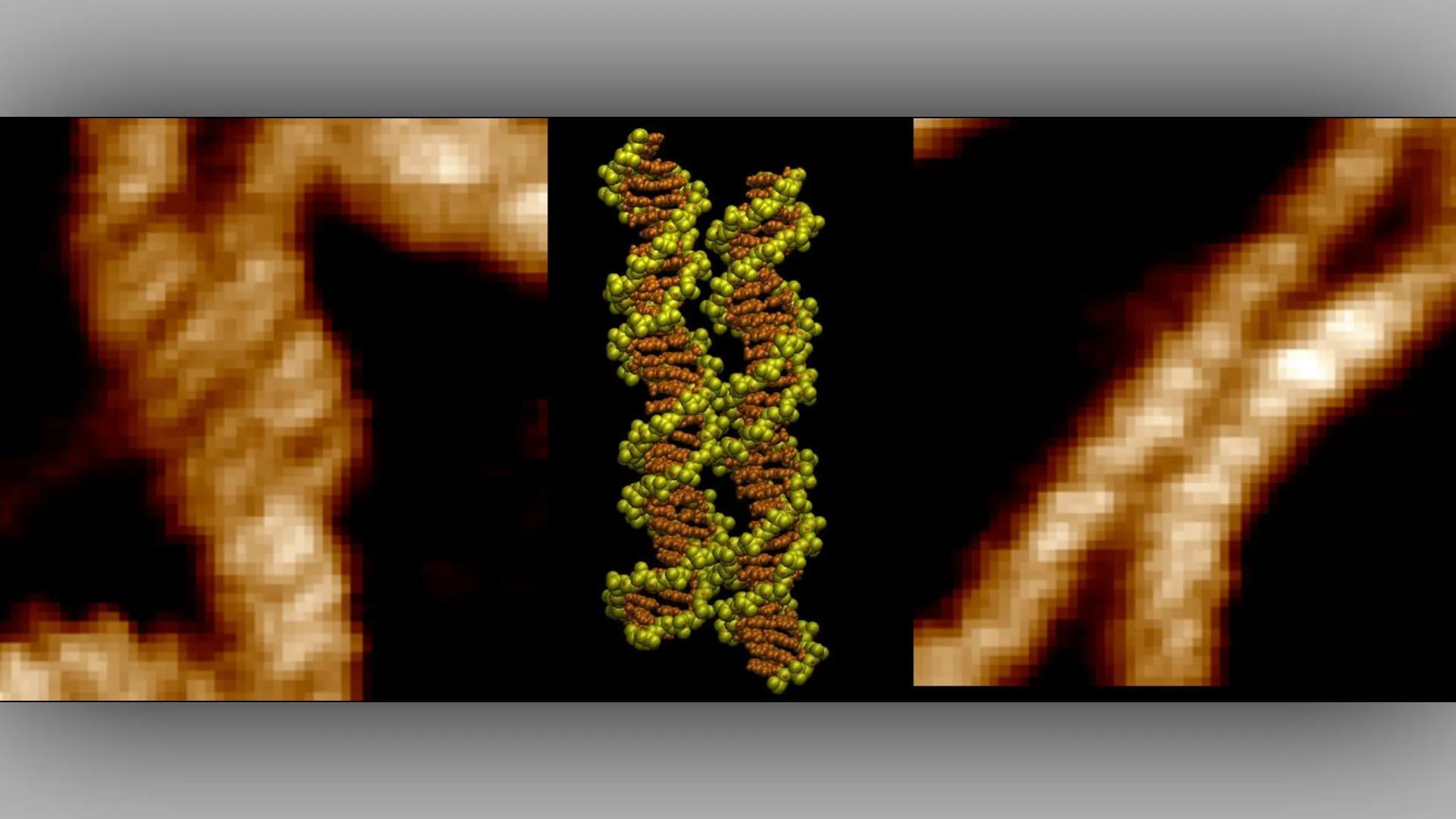
The researchers began by utilizing atomic force microscopy (AFM). Unlike traditional light microscopy, which is limited by the wavelength of visible light, AFM uses a physical probe to "feel" the surface of molecules, mapping their topography at the nanometer scale. By scanning DNA samples, the team was able to capture the physical positioning of the strands, providing the first visual evidence of DNA molecules aligning groove-to-groove with remarkable precision.
Phase 2: Simulating the Invisible
While the AFM provided the "what," the researchers needed to understand the "how." Dr. Victor Velasco-Berrelleza of the University of Sheffield took on the task of building sophisticated computer simulations. By modeling the movement of individual atoms and ions in real-time, the team observed how double-charged metal ions behaved. The simulations revealed that these ions do not simply float; they act as "charged arms" that reach out, docking into the grooves of two adjacent DNA strands simultaneously. This effectively neutralizes the repulsion and creates a stable, bridge-like connection that pulls the helices into alignment.
Phase 3: Synthesizing the Data
The final stage of the research involved reconciling the visual maps from the AFM with the molecular dynamics observed in the simulations. This integration allowed the team to confirm that the observed alignment in the physical samples was directly caused by the ion-bridging mechanism predicted by the Kornyshev model.
Unlocking the Mechanism: The Role of Divalent Ions
The study’s most profound finding is the specific behavior of divalent metal ions. In a cellular solution, these ions are ubiquitous. However, the study demonstrates that they are not merely passive spectators in the cell’s chemical soup.
The simulations showed that a single ion can interact with both DNA molecules at once. By positioning itself between the two helices, the ion acts as a temporary adhesive. This process is highly sequence-dependent. The team discovered that DNA does not pair with equal affinity across every region of the genome. Instead, there are distinct "hotspots"—specific sequences that create stronger contacts than others.
These hotspots are characterized by their ability to facilitate the perfect fit for the metal ion bridges. When a divalent ion settles into these grooves, it effectively "zips" the DNA segments together. This discovery explains why certain parts of the genome are more prone to recombination and why other parts remain tightly packed or shielded.
Official Responses and Scientific Impact
The research team expressed a shared sense of accomplishment in having finally witnessed a process that has been a pillar of theoretical biophysics for twenty years.
"It was incredible to be able to directly visualize the long-hypothesized mechanism for the first time," said Dr. Thomas Catley. "The advanced imaging techniques at our disposal are allowing us to uncover these key DNA interactions, which have implications in many key cellular processes."
Professor Agnes Noy emphasized the clinical relevance of the findings. "This discovery could help researchers identify regions of the genome specially involved in DNA pairing," she noted. "These regions may become particularly important when mutations disrupt normal cellular processes and contribute to cancer."
The importance of this work is not just in confirming a theory, but in providing a roadmap for future investigation. As Dr. Victor Velasco-Berrelleza explained, "Microscopy shows us what happens, but the simulations allow us to uncover the molecular mechanism behind it." By bridging the gap between imaging and simulation, the team has created a template for how scientists might study other complex molecular interactions in the future.
Implications for Oncology and Biotechnology
The identification of DNA "pairing hotspots" has far-reaching consequences, particularly in the field of oncology. Cancer is, at its core, a disease of genomic instability. When the mechanisms that control how DNA repairs itself or recombines go awry, the result can be the uncontrolled cellular growth characteristic of malignancy.
By mapping these hotspots, researchers may be able to better understand why certain regions of the genome are more susceptible to the mutations that drive cancer. If a specific region of DNA relies heavily on ion-mediated bridging for its stability, a disruption in the availability of those ions—or a mutation that alters the groove shape—could lead to genomic "fraying" or incorrect pairing, potentially triggering the onset of disease.
Beyond medicine, the discovery offers a new toolkit for biotechnology. The ability to program DNA to interact with specific affinity suggests a future where scientists can design "smart" DNA nanostructures. By engineering sequences that utilize ion-bridging to lock into place, researchers could construct complex, self-assembling biological machines, drug-delivery vehicles, or ultra-dense data storage devices made of synthetic DNA.
Future Directions: Beyond the Test Tube
While the current study marks a significant victory for structural biology, the researchers acknowledge that there is much work left to do. The next phase of research will likely involve observing these processes in even more complex, crowded environments that mimic the interior of a living cell more closely than current in vitro models.
Furthermore, the team intends to investigate how different types of ions and environmental factors—such as changes in pH or the presence of DNA-binding proteins—might modulate the DNA-zipper effect. By understanding these variables, scientists hope to gain a comprehensive view of how the genome is organized and maintained.
The study, titled "Imaging and mechanism of DNA-DNA recognition mediated by divalent ions," stands as a testament to the power of combining classical physical theory with modern computational and imaging prowess. As we continue to zoom in on the fundamental building blocks of life, discoveries like this remind us that the complexity of the cell is not just in the sequence of our genes, but in the physical forces that allow those genes to dance together.