
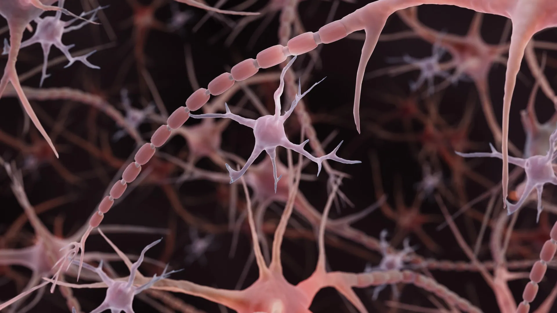
For over a century, the textbook illustration of a neuron has remained largely unchanged: a central cell body with spindly, smooth-tubed extensions known as axons, acting as the high-speed fiber-optic cables of the biological world. These axons, responsible for transmitting electrical impulses across the vast, intricate network of the human brain, have long been depicted as uniform, linear conduits.
However, a groundbreaking paradigm shift in neuroscience is currently underway. Recent research, spearheaded by scientists at Johns Hopkins Medicine, has revealed that these "cables" are not smooth at all. Instead, many axons resemble strings of tiny pearls, characterized by repeating, nanoscale bulges. These structures, far from being mere architectural irregularities, appear to be dynamic, functional components that actively regulate the speed and efficiency of neural communication.
The Discovery: Challenging a Century of Dogma
The traditional view of the axon as a rigid, uniform tube has been the foundation of neuroscience since the era of Santiago Ramón y Cajal. While scientists have long observed "beaded" axons, these were almost exclusively associated with pathological states—dying neurons, the ravages of neurodegenerative diseases like Parkinson’s, or the result of traumatic injury. In these contexts, beading is interpreted as a sign of membrane disintegration and structural failure.
In a study published in Nature Neuroscience on December 2, 2024, researchers at Johns Hopkins challenged this binary view. By utilizing high-pressure freezing—a technique that preserves cellular architecture in a state closer to life than traditional chemical fixation—the team identified "non-synaptic varicosities." Unlike the pathological swellings seen in disease, these nanoscale pearls were found in healthy, functioning neurons.
"To see nanoscale structures with standard electron microscopy, we fix and dehydrate the tissues, but freezing them retains their shape—similar to freezing a grape rather than dehydrating it into a raisin," explained Shigeki Watanabe, Ph.D., an associate professor of cell biology and neuroscience at the Johns Hopkins University School of Medicine. This distinction is vital: it suggests that pearling is not a sign of cellular death, but rather an integral, normal feature of healthy brain architecture.
A Chronology of Discovery
The journey toward redefining axonal anatomy was neither linear nor solitary. It began with an unlikely observation in a different species.
- Early Observations (Pre-2024): Watanabe’s interest was piqued by reports of repeating structures along the axons of C. elegans (worms). Initial hypotheses suggested that these pearls were formed by the axon’s internal protein cytoskeleton.
- Testing the Cytoskeleton Hypothesis: Working with graduate student Jacqueline Griswold, Watanabe collaborated with Swiss scientist Graham Knott to determine if the internal protein scaffold was the primary architect of these pearls. When the team disrupted the cytoskeleton, however, the pearling persisted, forcing them to look elsewhere.
- The Physics Turn: The team pivoted toward the biophysics of the cellular membrane. By partnering with theoretical biophysicist Padmini Rangamani of the University of California San Diego, they sought to determine if mechanical forces—such as membrane tension and composition—could account for the shape.
- The 2024 Breakthrough: The Johns Hopkins study confirmed the presence of these structures in mouse neurons across various stages of development. Using electron microscopy, they validated that these shapes were present in vivo, ruling out artifacts caused by laboratory processing.
- Human Translation (2025–2026): In November 2025, a study published in Neuron led by Chelsy R. Eddings and Dr. Watanabe confirmed that these pearled axons are not limited to rodent models. By analyzing human cortical tissue obtained during epilepsy surgery, the team demonstrated that these structures exist within the human brain, bridging the gap between theoretical models and human clinical relevance.
The Physics of Signaling: How Form Dictates Function
If the axon is a cable, the pearled structure acts as a series of physical "valves" or "tunnels." The Johns Hopkins team discovered that the dimensions of these varicosities—and the narrow gaps between them—directly influence the velocity of electrical signals.
Through mathematical modeling, the researchers found that membrane tension and composition, specifically the presence of cholesterol, play a critical role. Removing cholesterol rendered the membrane more fluid and less stiff, which in turn altered the pearled geometry and slowed the transmission of electrical impulses.
"A wider space in the axons allows ions to pass through more quickly and avoid traffic jams," Watanabe noted. This implies that the brain can "tune" its transmission speeds by subtly adjusting the physical diameter of these pearls. This discovery suggests that the brain is not merely a collection of fixed wires, but a fluid, responsive system where physical shape is a primary regulator of information processing.
Neural Plasticity: The Dynamic Brain
Perhaps the most startling finding is that these pearls are not static. The Johns Hopkins team subjected neurons to high-frequency electrical stimulation—simulating the intense activity of a brain processing information or learning a new skill. Following this stimulation, the pearl-like regions underwent measurable changes, becoming significantly longer and wider.
This enlargement persisted for at least 30 minutes, during which time the speed of electrical signal propagation decreased. This suggests that the axon’s structure is intrinsically linked to its recent activity history. By changing its shape, the axon may be providing a mechanism for "synaptic scaling" or temporary modulation of signal strength, potentially playing a role in how the brain manages the transition from short-term memory to long-term consolidation.
Implications for Neurological Health and Disease
The existence of these pearls in healthy tissue necessitates a complete re-evaluation of how we interpret diagnostic brain imaging. For decades, clinicians have viewed "beading" in neural tissue as an unambiguous marker of damage. If, however, healthy axons naturally exist in a pearled state, the line between physiological function and pathological decay becomes blurred.
The research team is now focused on the crucial distinction between "normal" pearling and "disease-associated" beading. Understanding the biophysical triggers that cause an axon to transition from a healthy pearl to a pathological, damaged state could open new avenues for treating neurodegenerative conditions like Alzheimer’s, Parkinson’s, and Amyotrophic Lateral Sclerosis (ALS). If we can stabilize the healthy, functional membrane structure, we may be able to prevent the catastrophic failure of neurons in these diseases.
Future Research Directions
The field is moving quickly. With a National Institute of Mental Health (NIMH) grant, Watanabe and Rangamani are expanding their research into complex computational models that simulate how incoming signals influence axonal shape in real-time.
Furthermore, the search for pharmacological control is underway. A July 2026 study in PLOS Biology by researchers at the University of Edinburgh utilized high-throughput screening on zebrafish to identify 33 chemical compounds capable of altering axon diameter. While these findings do not directly replicate the pearling phenomenon, they provide a powerful toolkit for researchers to manipulate neuronal dimensions, potentially offering a way to "rescue" axons that have lost their optimal signaling shape.
Conclusion: A New Frontier in Neuroscience
The discovery that the brain’s "cables" are dynamic, pearl-like structures represents a fundamental shift in our understanding of the nervous system. By moving beyond the static, smooth-tube model, scientists are uncovering a level of complexity previously hidden by the limitations of traditional imaging.
The brain, it appears, is far more flexible than we ever imagined—both in its signaling speed and its structural capacity to adapt to experience. As researchers continue to map the interplay between membrane physics and electrical activity, they are not just rewriting textbooks; they are uncovering the physical mechanisms of thought, memory, and perhaps, the very nature of human consciousness. The "pearls" in our brain, once dismissed as noise, are proving to be the silent orchestrators of our most complex cognitive functions.