
Long before a human infant draws its first breath, the foundation of their future—the cerebral cortex—is meticulously constructed through a series of complex cellular mandates. At the heart of this biological masterpiece lies the radial glia, a specialized class of neural stem cells that act as the master architects of the human brain. Responsible for the genesis of the cerebral cortex—the region governing complex thought, memory, and language—these cells are fundamentally what set humans apart from other species.
New research from the David Geffen School of Medicine at UCLA, published in the prestigious journals Cell and Science, has unveiled that these architects do not work in a vacuum. Instead, radial glia are subject to a constant, sophisticated dialogue with their environment, guided by metabolic signals and physical contact from distant brain regions.
The Architectural Blueprint: The Role of Radial Glia
Radial glia are arguably the most influential cells in the human body. They serve as the primary source for the vast array of neurons and support cells that constitute the cerebral cortex. Beyond their role in basic development, they are believed to be the primary drivers behind the extraordinary expansion of the human cortex compared to that of other primates and rodents.
While the majority of these cells complete their life cycle and disappear before birth, their legacy remains imprinted in the architecture of the brain. Intriguingly, similar cellular programs have been observed to re-emerge in brain cancers, suggesting that the "decision-making" machinery of these cells is not only vital for development but potentially prone to corruption.
"Radial glia are the coolest cells that have ever existed," says Dr. Aparna Bhaduri, assistant professor of biological chemistry at the David Geffen School of Medicine at UCLA. "They are really key to making us human. But they are also at the center of many neurodevelopmental and neuropsychiatric disorders, as well as cancer—so understanding how they make their decisions is one way to start understanding how those conditions arise."
Chronology of Discovery: From Metabolism to Physical Touch
The journey to these findings involved a multi-year effort to map the hidden variables influencing stem cell behavior. The research trajectory can be viewed through two distinct lenses: the metabolic environment and the physical architecture of early neural communication.
Phase 1: The Metabolic Atlas (Published in Cell)
In collaboration with the lab of Heather Christofk, researchers led by Jessenya Mil and Jose Soto set out to determine if metabolism was merely a "background" process providing fuel, or an active signaling system. By analyzing donated human tissue and brain organoids, the team constructed a high-resolution metabolic atlas of the developing cortex.
They discovered that radial glia rely heavily on the pentose phosphate pathway. When the team throttled glucose availability or disrupted this pathway, the stem cells’ output shifted. They began producing inhibitory neurons—cells that usually appear much later in the developmental sequence—rather than the standard excitatory cells. This proved that metabolism serves as a regulatory switch for cell identity.
Phase 2: The Thalamic Connection (Published in Science)
Led by Claudia Nguyen, the second study investigated the "why" behind the early arrival of thalamic fibers. It has long been established that the thalamus, a deep-brain relay station, sends fibers toward the cortex. However, in humans, these fibers arrive well before their final connections are functional.
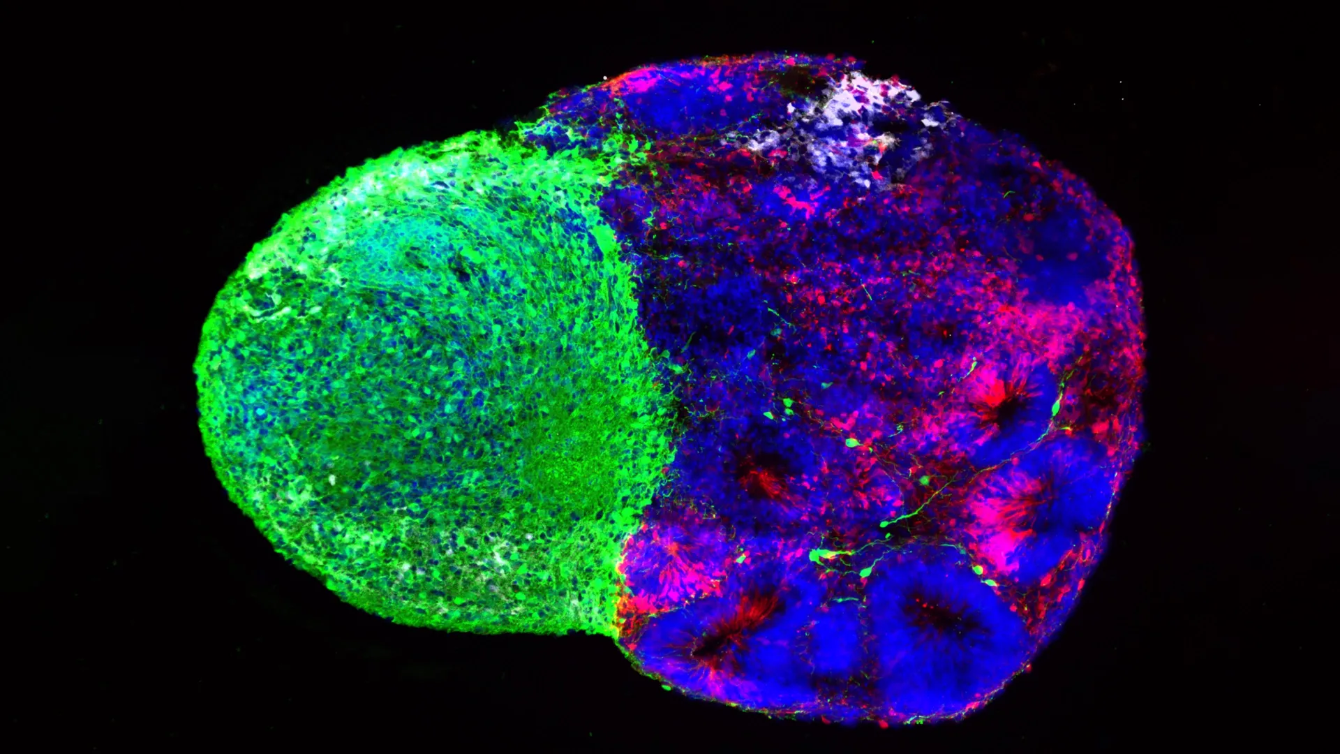
The UCLA team utilized "assembloids"—complex, 3D laboratory models that fuse different brain tissues—to observe these fibers in action. They discovered that thalamic projections physically touch radial glia, triggering them to produce excitatory neurons, specifically those in the upper layers of the cortex. This physical contact appears to be a uniquely human evolutionary development, absent in simpler animal models.
Supporting Data: The Mechanics of Decision-Making
The evidence gathered by the UCLA team challenges the traditional view of neural development as a purely genetic, hard-coded program.
- Metabolic Influence: The study demonstrates that radial glia act as metabolic sensors. By prioritizing the pentose phosphate pathway, the cells can maximize the production of materials necessary for rapid division. When this process is perturbed, the "birth order" of neurons is disrupted, leading to a potential cascade of developmental issues.
- The NRXN1 Factor: The research connected these findings to the gene NRXN1, which is heavily implicated in autism spectrum disorder. By creating assembloids using cells from patients with NRXN1 mutations, the team observed that the thalamic signals were altered. This suggests that the physical interaction between the thalamus and the cortex is a critical window of vulnerability; if the signal or the response is "noisy" due to genetic mutations, the fundamental composition of the cortex is altered.
Official Responses and Scientific Perspective
The implications of these studies reach far beyond basic neurobiology. For researchers in the field, these findings represent a paradigm shift in how to interpret brain development.
Dr. Bhaduri, who is also a member of the UCLA Broad Stem Cell Research Center and the UCLA Health Jonsson Comprehensive Cancer Center, emphasizes that the scientific community must look at the brain’s "operating system" with more nuance. "What was surprising is that metabolism isn’t just a passive thing that happens in the background," she states. "It can really control how stem cells make decisions."
The research also highlights the evolution of organoid technology. A decade ago, scientists were limited to observing static samples or relying on mouse models that failed to capture the nuances of human cortical expansion. Today, the ability to engineer assembloids—miniature, functional "brains-in-a-dish"—allows for the testing of hypotheses that were previously relegated to the realm of theory.
Broader Implications: A Future of Precision Medicine
The convergence of these two studies points toward a unified theory: the developing brain is in a state of constant, high-stakes communication. The radial glia act as a central processing unit, integrating metabolic "fuel" data with physical "structural" data to decide whether to continue expanding the stem cell pool or to begin differentiating into specialized neurons.
Impact on Clinical Research
- Environmental and Maternal Health: The discovery that metabolism dictates cell fate suggests that maternal nutrition and metabolic health during pregnancy could have a direct, measurable impact on the cortical architecture of the fetus. This provides a clear, biological pathway to investigate how environmental stressors impact neurodevelopment.
- Neuropsychiatric Disorders: By linking NRXN1 mutations to the physical interaction between the thalamus and the cortex, researchers now have a specific target for study. This could lead to earlier diagnostic markers or potential interventions for conditions like autism and schizophrenia, which are rooted in early developmental failures.
- Oncology: As radial glia behavior is often hijacked by brain cancers, understanding the decision-making "switches" of these stem cells offers a potential roadmap for cancer therapies. If scientists can force cancer cells to revert to a less aggressive state or cease division by manipulating these metabolic pathways, it could open a new frontier in pediatric and adult brain cancer treatment.
Conclusion: Looking Under the Hood
The research led by the UCLA team provides a profound glimpse "under the hood" of human development. By proving that radial glia are active participants in their own environment—rather than static responders to genetic code—the scientists have unlocked new ways to view the human brain.
"Ultimately, these studies give us a glimpse under the hood of how these cells make decisions," Bhaduri concludes. "Understanding those decisions is a first step toward understanding normal brain development, disease vulnerability, and, potentially, how similar stem-cell programs operate in brain cancer."
As organoid technology continues to mature, the ability to study the human brain in real-time offers a hopeful future. The days of treating the brain as a black box are coming to an end; in their place, we are building a precise, evidence-based map of the most complex structure in the known universe.
This research was supported by a wide coalition of organizations, including the National Institutes of Health, the National Science Foundation, the Brain & Behavior Research Foundation, the Alfred P. Sloan Foundation, the Rose Hills Foundation, the Esther A. & Joseph Klingenstein Fund, the Simons Foundation, the Chan Zuckerberg Initiative, the NIH BRAIN Initiative Cell Atlas Network, the International Foundation for Ethical Research, the UCLA Broad Stem Cell Research Center’s Stem Cell Research Training Program, and the UCLA Health Jonsson Comprehensive Cancer Center and UCLA Broad Stem Cell Research Center Ablon Scholars Program.