
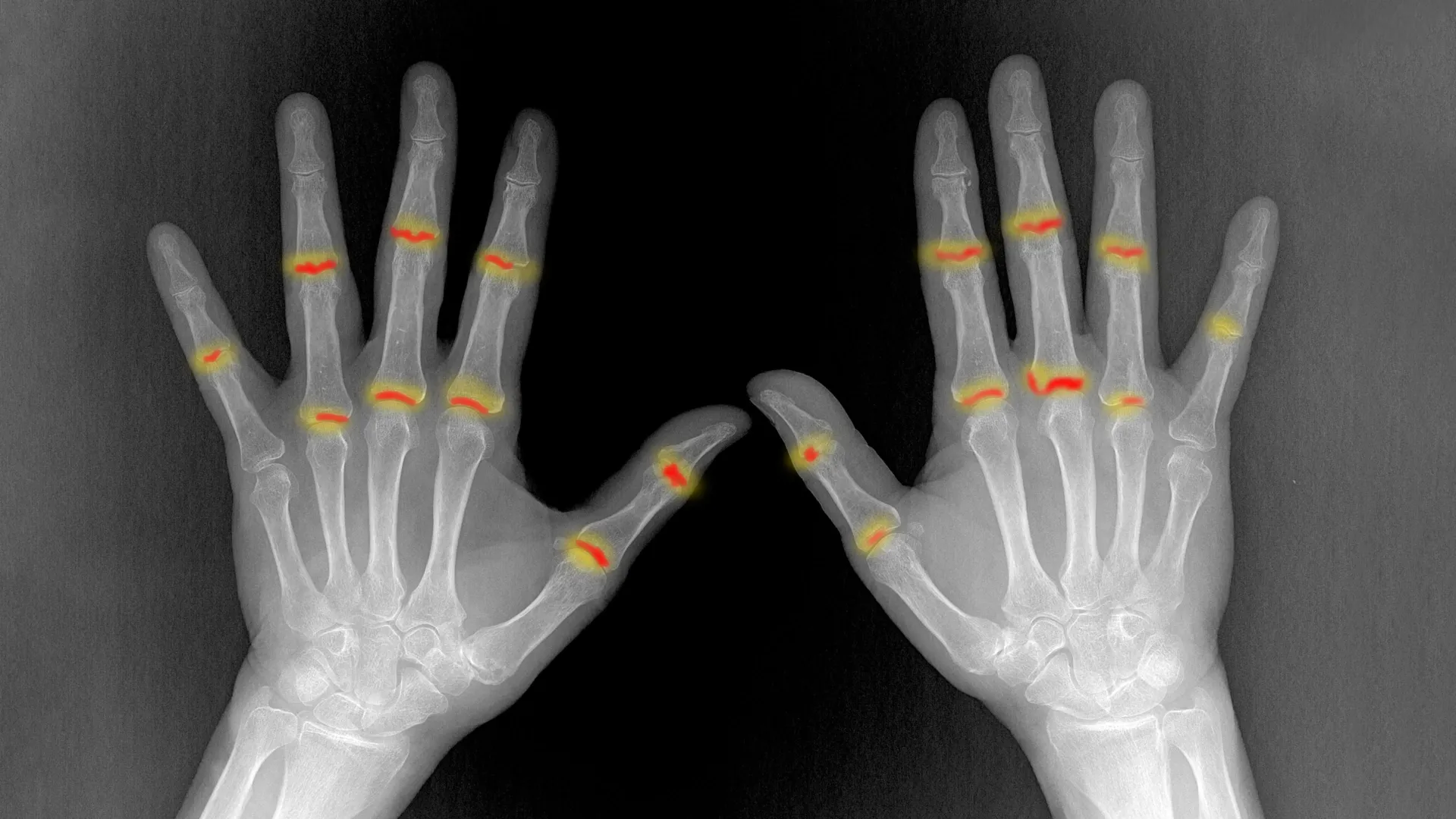
For decades, the medical community has grappled with a frustrating biological paradox: why does rheumatoid arthritis (RA)—a systemic autoimmune disease—choose to wreak havoc on specific joints while leaving others, often mere millimeters away, completely unscathed? This selectivity has long been a defining mystery of the condition, which affects millions worldwide, causing chronic pain, stiffness, and permanent joint destruction.
A groundbreaking study published in Nature Immunology titled "The embryonic origins of site-specific arthritis" has finally provided a compelling answer. Researchers at the Kennedy Institute of Rheumatology at the University of Oxford have discovered that the susceptibility of a joint to inflammation is not solely determined by the immune system in adulthood. Instead, it is written into the genetic and structural "blueprint" of the joint long before a person is even born.
The Mystery of Selective Vulnerability
Rheumatoid arthritis is characterized by an immune-mediated attack on the synovium, the delicate, thin tissue lining the joints. When this lining becomes inflamed, it triggers a cascade of damage that can erode cartilage and bone. While the immune system’s role is well-documented, the "geography" of the disease—its tendency to target the proximal interphalangeal (PIP) joints of the fingers while sparing the distal interphalangeal (DIP) joints—has remained poorly understood.
By comparing these two types of finger joints, which sit side-by-side but react differently to the disease, the Oxford-led team sought to determine if the local tissue environment acts as a "gatekeeper" for inflammation. Their findings suggest that the body’s vulnerability to autoimmune attacks is deeply rooted in developmental biology.
Chronology of the Discovery
The research journey began with a shift in perspective. Rather than focusing exclusively on the diseased adult joint, researchers turned their attention to the earliest stages of human skeletal development.
- Mapping the Embryo: Using cutting-edge single-cell sequencing and high-resolution 3D X-ray imaging, the team mapped the development of human finger joints in utero. This allowed them to capture the "formative years" of the joint, a window of time where cell identities are established.
- Identifying Cellular Signatures: During this phase, they observed that developing joints are dominated by structural cells—specifically fibroblasts and chondrocytes (cartilage-forming cells)—rather than immune cells.
- Detecting Pre-existing Differences: As the joints took shape, the team identified significant discrepancies between the PIP and DIP joints. These structural differences, including the volume of synovial tissue and the density of specific fibroblast populations, were already present in the fetal stage.
- Validating the "Trigger" Mechanism: By exposing these cells to inflammatory signals in a laboratory setting, the researchers observed that the fibroblasts from vulnerable joints (PIP) responded with a distinct, heightened inflammatory profile compared to those from "spared" joints (DIP).
Supporting Data: The Role of PI16+ Fibroblasts
The core of the study rests on the identification of a specific cell type: PI16-positive (PI16+) fibroblasts. These specialized connective tissue cells were found in significantly higher concentrations within the joints prone to rheumatoid arthritis.
Structural and Biological Divergence
The researchers employed a custom-built image analysis tool to visualize the spatial distribution of these cells. They found that PI16+ fibroblasts are not randomly distributed; they cluster around blood vessels and at the critical junctions where tendons and ligaments anchor to surrounding tissue.
This location is significant. These anchor points are areas of high mechanical stress, and the presence of these specific cells may be an evolutionary adaptation to support joint movement. However, in the context of autoimmune disease, these cells appear to be "primed" for a maladaptive response.
The data showed that when challenged with inflammatory stimuli, PI16+ fibroblasts exhibited a unique biological pathway. While all fibroblasts in the joint can contribute to inflammation, the PI16+ population showed distinct changes in immune regulation and tissue organization genes. This suggests that these cells act as "sentinels" that, when triggered, facilitate the recruitment of immune cells, effectively opening the door for the disease to take hold.
The 3D Perspective
Using the advanced capabilities of the Diamond Light Source—the UK’s national synchrotron science facility—the team created high-resolution 3D maps of the joints. These images confirmed that it is not just the cells, but the architecture of the synovium itself that differs. Vulnerable joints possess a more complex, voluminous synovial lining, which provides a larger "playing field" for inflammatory cells to proliferate once the immune system begins its attack.
Official Responses and Expert Insight
The implications of this study are profound, marking a departure from the "immune-centric" view of autoimmune disease.
Christopher Buckley, Kennedy Professor of Translational Rheumatology at the University of Oxford and the study’s senior author, emphasized the shift in scientific understanding. "For decades we have known that rheumatoid arthritis selectively targets particular joints, but one of the great unanswered questions is why? Our findings suggest that the answer lies not only in the immune system but also in the tissues themselves. The cellular and structural characteristics established during development may help determine where inflammation takes hold later in life."
Dr. Sarah Davidson, a postdoctoral researcher at the Kennedy Institute and a first author of the study, highlighted the importance of the embryonic window. "We found that joints commonly affected by rheumatoid arthritis already contain distinct cellular populations before birth. PI16+ fibroblasts were enriched in vulnerable joints and responded differently to inflammatory signals. Their location and behavior suggest they could help shape where disease develops."
The research team, which included collaborators from the University of Birmingham, University College London, and the Diamond Light Source, has effectively proven that the "soil" (the joint tissue) is just as important as the "seed" (the immune system’s overreaction) in the development of arthritis.
Implications for Future Medicine
The discovery that RA vulnerability is "pre-programmed" in the joint’s development opens several new avenues for treatment and prevention.
1. Precision Therapeutics
Current treatments for rheumatoid arthritis often involve systemic immunosuppression, which can have significant side effects. If scientists can identify the specific molecular pathways that make PI16+ fibroblasts hyper-responsive to inflammation, they may be able to develop targeted therapies that "calm" these cells without suppressing the entire immune system.
2. Regenerative Medicine
Understanding the developmental signals that lead to the creation of the synovial lining offers clues for regenerative medicine. If researchers can decode these signals, they may one day be able to "reprogram" or restore the normal, protective behavior of synovial fibroblasts, potentially reversing the damage caused by chronic inflammation.
3. Early Intervention and Risk Assessment
While the study does not suggest that arthritis is purely genetic, it does suggest that an individual’s joint architecture could serve as a biomarker for disease risk. Future diagnostic tools could potentially use high-resolution imaging to assess an individual’s joint structure, helping doctors predict which patients are at higher risk for severe joint involvement and allowing for earlier, more aggressive preventative care.
Conclusion: A New Era in Rheumatology
The work conducted at the Kennedy Institute serves as a bridge between developmental biology and clinical rheumatology. By demonstrating that the susceptibility to rheumatoid arthritis is a life-long trait established in the womb, the study fundamentally changes how we view the disease.
We are no longer looking at RA as a simple failure of the immune system. Instead, we are seeing it as an interaction between a systemic immune response and a pre-existing local tissue landscape. As researchers continue to map the cellular architecture of human joints, the hope is that these "developmental clues" will lead to a new generation of therapies—treatments that don’t just manage the symptoms of arthritis, but address the very foundation of why it targets the body in the way it does.
This research, supported by the Medical Research Council, is a testament to the power of high-resolution, interdisciplinary science in solving medical mysteries that have baffled clinicians for generations. The blueprint of the joint is no longer just a structural map; it is now a roadmap for the future of autoimmune research.