
In a breakthrough that could redefine the clinical management of joint trauma, researchers at The University of Alabama in Huntsville (UAH)—a prominent institution within The University of Alabama System—have identified a novel, drug-free application for continuous low-intensity ultrasound. The study, recently published in the prestigious Nature journal Scientific Reports, suggests that this non-invasive mechanical stimulation can effectively "reprogram" the body’s immune response, pivoting it from a destructive state of chronic inflammation toward a regenerative cycle of tissue repair.
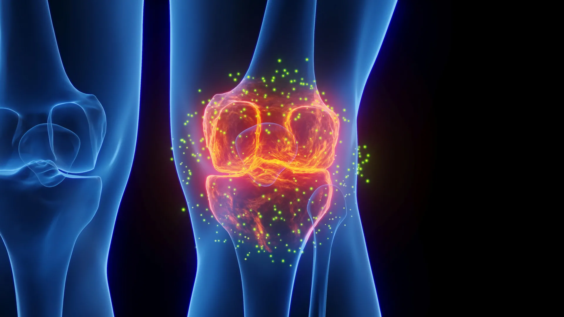
This discovery offers a potential lifeline to the millions of individuals who suffer from post-traumatic osteoarthritis (PTOA)—a degenerative condition often triggered by the body’s own failed attempts to heal following a severe joint injury. By leveraging the body’s inherent biological machinery rather than relying on pharmacological interventions, the UAH team has mapped a path toward a new frontier in regenerative medicine.
The Core Findings: Harnessing Mechanics to Modulate Biology
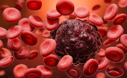
At the heart of the research lies the macrophage, a specialized white blood cell that acts as the immune system’s frontline responder. Following a joint injury, macrophages arrive in two primary forms: the inflammatory "defender" (M1) cells, designed to clear cellular debris and pathogens, and the regenerative "healer" (M2) cells, which orchestrate the complex process of tissue reconstruction.
Under normal circumstances, the body transitions seamlessly from the M1 phase to the M2 phase. However, in the case of severe joint trauma, this process often stalls. The immune system remains locked in a hyper-inflammatory M1 state, creating a toxic, acidic environment that damages healthy cartilage and accelerates the progression of osteoarthritis.
The UAH team, led by Dr. Anuradha Subramanian, professor of chemical and materials engineering, discovered that applying continuous low-intensity ultrasound can mechanically signal these macrophages to abandon their inflammatory stance. By exposing the cells to precise, low-intensity waves, the researchers observed a molecular shift that promotes the M2-like phenotype, effectively "rebooting" the immune system to favor recovery over destruction.
Chronology of the Investigation
The project, funded by a substantial R01 grant from the National Institutes of Health (NIH), represents years of cross-disciplinary collaboration. The development of the study can be segmented into three critical phases:
Phase I: Experimental Design and Biological Modeling
The study began with the work of Dr. Shahid Khan during his doctoral studies. Recognizing that traditional laboratory models often fail to replicate the complexity of an injured joint, the team pivoted to a more sophisticated approach. They utilized fibronectin fragments—molecules released naturally when tissues undergo mechanical stress and degradation. By introducing these fragments to the immune cells, the team created a "micro-environment" that mimicked the post-traumatic state of a human knee or shoulder more accurately than standard experimental protocols.
Phase II: Computational Analysis and Transcriptomics
Once the biological response was triggered, the challenge shifted to analyzing the massive amounts of data generated. Dr. Satyaki Roy, a professor of mathematical sciences at UAH, spearheaded the computational strategy. Rather than looking at individual gene expressions—a method that often misses the "big picture" of cellular behavior—the team employed "differential clustering." This advanced computational technique allows researchers to observe how large networks of genes move in concert. By mapping these gene clusters, the researchers could identify the precise biological "switches" being toggled by the ultrasound.
Phase III: Validation and Result Synthesis
In the final stages, the team—bolstered by the contributions of graduate student Owen Trippany—confirmed that the ultrasound-treated cells showed a marked decrease in inflammatory biomarkers. Simultaneously, they documented a robust uptick in markers associated with tissue remodeling and repair. This phase finalized the evidence that the intervention was not just stopping the "bad" inflammation but actively stimulating the "good" repair processes.
Supporting Data: Why This Approach Matters
The significance of this research is rooted in the clinical failure of current treatments for post-traumatic osteoarthritis. Currently, patients with joint injuries often rely on non-steroidal anti-inflammatory drugs (NSAIDs) or corticosteroids. While these can manage pain, they do little to address the root cause of the cellular "stuck" state that leads to long-term joint degeneration.
The Problem with Persistent Inflammation
As Dr. Satyaki Roy explains, the pathology of post-traumatic osteoarthritis is a feedback loop. "Post-traumatic osteoarthritis is driven in part by persistent inflammation that limits tissue repair and accelerates joint degeneration," Roy notes. When the M1 macrophages remain dominant, the inflammatory cytokines they release degrade the extracellular matrix of the cartilage. This, in turn, releases more fragments that stimulate more inflammation, creating a self-perpetuating cycle of damage.
The Ultrasound Advantage
The UAH team’s data suggests that continuous low-intensity ultrasound acts as a "mechanical interrupt" for this cycle. By providing a non-pharmacological stimulus, the researchers were able to influence cellular behavior without the systemic side effects associated with high-dose anti-inflammatory medications. The ability to shift macrophages toward an M2-like state is the "holy grail" of current immunomodulation research, as it holds the potential to preserve healthy cartilage while simultaneously accelerating the healing of torn ligaments or damaged menisci.
Official Perspectives: From the Lab Bench to the Clinic
The interdisciplinary nature of the UAH team allowed for a holistic view of the problem, blending the physical principles of ultrasound with the high-level data analysis of modern genomics.
Dr. Anuradha Subramanian: A Vision for Drug-Free Healing
Dr. Subramanian emphasizes the long-term potential of this technology. "In an ‘M1’ state, macrophages promote inflammation to fight damage or infection, but prolonged M1 activity can also harm healthy tissue," she states. "Our findings suggest that continuous low-intensity ultrasound may help restore this balance by promoting a more reparative macrophage response." She underscores that the ultimate goal is not to eliminate the inflammatory response entirely—which is necessary for initial injury cleanup—but to ensure that the body knows when to transition to the repair phase.
Dr. Satyaki Roy: The Power of Coordinated Data
Dr. Roy highlights the importance of the team’s computational methods in validating the treatment. "This allowed us to study not only which genes changed, but also how groups of genes changed their coordinated behavior in response to ultrasound stimulation," he says. This, he argues, provides a more reliable foundation for future clinical trials, as it proves that the effect is systemic across the cell’s genetic architecture rather than a minor, localized fluctuation.
Implications: A New Era for Orthopedic Care
The implications of the UAH study extend far beyond the laboratory walls. If validated in upcoming clinical trials, this technology could fundamentally change the trajectory of recovery for professional athletes, military personnel, and elderly patients prone to falls.
The Path Forward: Animal Models and Beyond
The team is already looking toward the next phase of development. "The next steps will involve validating these findings in animal models of early post-traumatic osteoarthritis and studying how ultrasound-based modulation affects long-term tissue repair in joint injury settings," Dr. Subramanian explains. These in vivo studies will be critical to understanding how the depth of the joint and the nature of the specific tissue (cartilage vs. synovial fluid) impact the efficacy of the ultrasound waves.
Potential for Clinical Integration
Because ultrasound is already a widely used, safe, and relatively inexpensive diagnostic tool, the infrastructure for this therapy already exists. Unlike complex new pharmaceuticals that require decades of safety testing and high manufacturing costs, the hardware required for this treatment is well-understood. If the UAH team can successfully optimize the intensity and duration of the ultrasound application, it could be integrated into standard physical therapy regimens, allowing patients to receive "immune-modulating" treatments during their regular recovery sessions.
Reducing the Burden on Healthcare Systems
The economic and societal impact of osteoarthritis is staggering. Millions of joint replacement surgeries are performed annually, many of which are the direct result of untreated or poorly healed trauma. By intervening early with a non-invasive, cost-effective strategy, this research could potentially delay or even prevent the need for invasive surgeries, drastically reducing the economic burden on healthcare systems and improving the quality of life for millions.
Conclusion
The research conducted at The University of Alabama in Huntsville represents a masterclass in modern scientific inquiry: identifying a biological bottleneck, applying rigorous interdisciplinary methodology to understand it, and finding a mechanical solution that respects the body’s natural rhythm. By proving that sound waves can influence the behavior of immune cells, the UAH team has opened a window into a future where healing is guided, not just managed.
As the team moves toward animal trials and eventually human clinical studies, the scientific community will be watching closely. If the promise of this study holds true, we may be on the verge of a new standard of care—one where the sound of healing is not just a metaphor, but a literal, measurable, and highly effective therapeutic reality.