
In a breakthrough that fundamentally alters our understanding of human ocular development, researchers at Johns Hopkins University have identified the precise biological mechanism that grants humans the ability to perceive the world with razor-sharp clarity. By studying lab-grown retinal tissue, the team has uncovered a sophisticated, time-sensitive "molecular handoff" between vitamin A derivatives and thyroid hormones that dictates the formation of the foveola—the tiny, critical center of the retina.
This discovery not only solves a decades-old mystery in developmental biology but also provides a vital roadmap for regenerative medicine. By clarifying how the eye constructs its most sensitive region, researchers are laying the groundwork for future cell-replacement therapies aimed at curing currently irreversible conditions such as macular degeneration and glaucoma.
The Architecture of Sight: Why the Foveola Matters
To understand the magnitude of this discovery, one must appreciate the anatomy of the human eye. While the human retina is a complex, multi-layered structure, the foveola—a minuscule pit at the very center—punches far above its weight. Though it occupies only a tiny fraction of the total retinal surface area, it is responsible for approximately half of all human visual perception.
The foveola is densely packed with cone photoreceptors, the specialized cells that allow for high-acuity daytime and color vision. Unlike the peripheral retina, which contains a mosaic of blue, green, and red cones, the foveola is exclusively populated by red and green cones. This specific configuration is what allows humans to read, recognize faces, and perform tasks requiring fine detail. For decades, the biological "blueprint" that ensures this specific cellular arrangement remained elusive, largely because the process cannot be adequately studied in common laboratory models like mice or zebrafish, which lack a foveola entirely.
Chronology: The Timeline of a Cellular Transformation
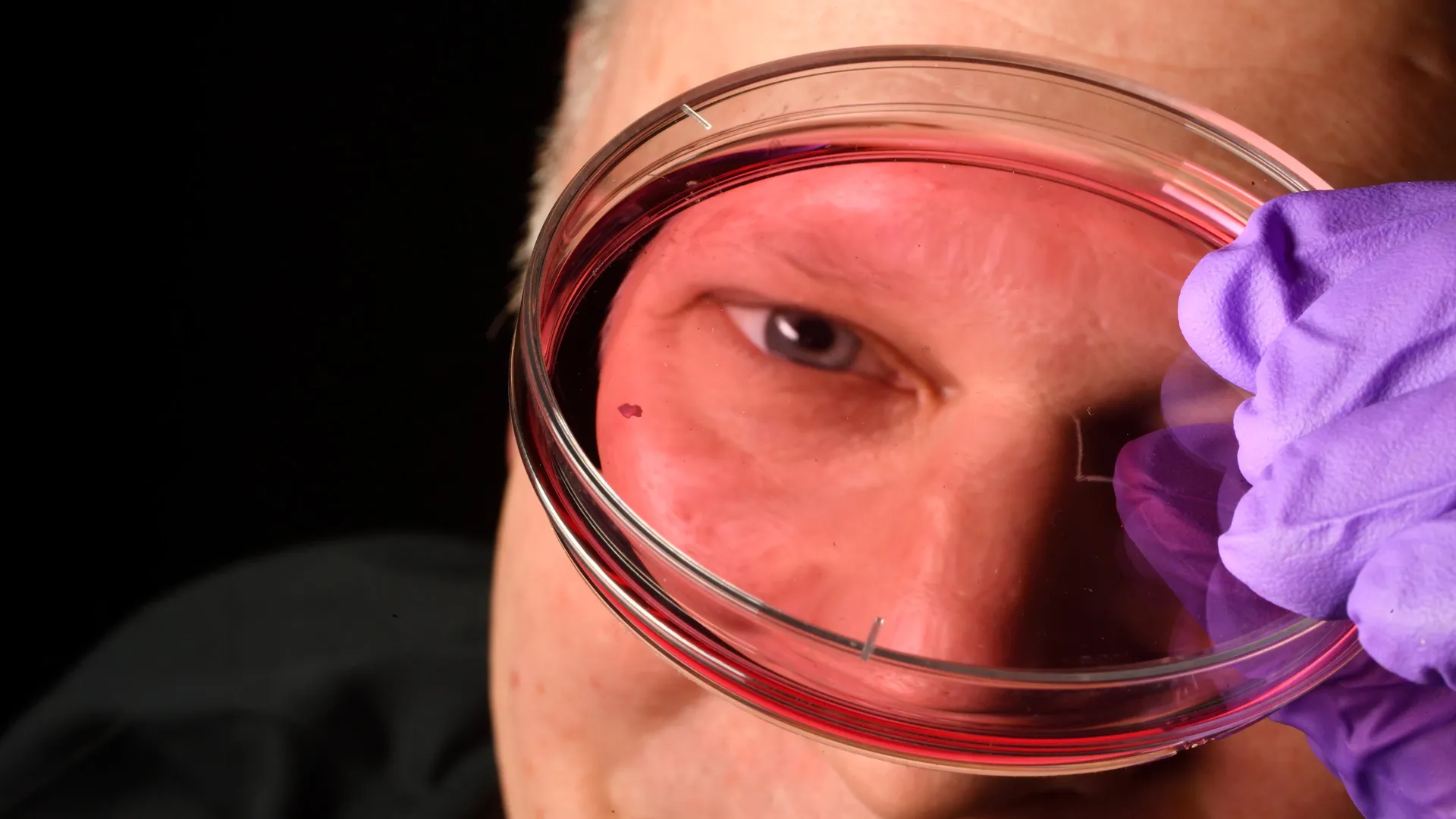
The research, published in the Proceedings of the National Academy of Sciences, relied on advanced organoid technology—small, three-dimensional clusters of tissue grown from fetal stem cells that mimic the structure and function of the human retina. By tracking these organoids over several months, the Johns Hopkins team was able to witness the fetal development of the eye in real-time.
The findings reveal a highly choreographed sequence of events occurring between the 10th and 14th weeks of gestation:
- Weeks 10–12: During this early window, the developing foveola begins to produce a small, initial population of blue-light-sensitive cones. Under normal circumstances, this would lead to a mixed population of cones.
- The First Mechanism (Retinoic Acid): As development progresses, the concentration of retinoic acid—a molecule derived from vitamin A—is strictly regulated and broken down. This degradation acts as a "stop" signal, curbing the formation of any additional blue cones.
- The Second Mechanism (Thyroid Hormone): Following the depletion of retinoic acid, thyroid hormones enter the biological scene. These hormones act as a transformative agent, signaling the remaining blue-sensitive cells to undergo a phenotypic shift, converting them into red or green cones.
By the 14th week, the transition is complete. The blue cones that were initially present have been effectively "swapped out" or converted, leaving the foveola with the precise red-green composition required for high-resolution vision.
Challenging a Longstanding Scientific Dogma
For over 30 years, the prevailing theory in ophthalmology was based on the concept of cell migration. Scientists previously believed that the blue cones formed in the center of the retina and were subsequently "pushed out" to the periphery as the eye developed, allowing red and green cones to take their place in the center.
The Johns Hopkins team’s evidence provides a radical alternative: the cells do not move; they change their identity.
"The main model in the field from about 30 years ago was that somehow the few blue cones you get in that region just move out of the way," explains Robert J. Johnston Jr., an associate professor of biology at Johns Hopkins who led the research. "We can’t really rule that out entirely yet, but our data supports a different model. These cells actually convert over time, which is really surprising."
By identifying that cell conversion—rather than physical migration—is the primary driver of foveal composition, the researchers have corrected a fundamental misconception, opening new avenues for how scientists might manipulate or "reprogram" cells in a therapeutic setting.
Official Responses and Expert Perspective
The implications of this study are being felt across the scientific community. By bridging the gap between fetal development and adult pathology, the team has provided a new "master switch" that researchers can target.
"This is a key step toward understanding the inner workings of the center of the retina, a critical part of the eye and the first to fail in people with macular degeneration," says Professor Johnston. "By better understanding this region and developing organoids that mimic its function, we hope to one day grow and transplant these tissues to restore vision."
Dr. Kimberly Hussey, a molecular and cell biologist formerly of the Johns Hopkins team and now with the Chicago-based cell therapy company CiRC Biosciences, emphasizes that the journey from the petri dish to the clinic is a marathon, not a sprint.
"The goal with using this organoid tech is to eventually make an almost made-to-order population of photoreceptors," Hussey noted. "A big avenue of potential is cell replacement therapy to introduce healthy cells that can reintegrate into the eye and potentially restore that lost vision. These are very long-term experiments, and of course, we’d need to do optimizations for safety and efficacy studies prior to moving into the clinic. But it’s a viable journey."
Implications: The Future of Vision Restoration
The clinical implications of these findings are profound. Diseases like age-related macular degeneration (AMD) are characterized by the progressive death of photoreceptors in the macula—the area that includes the foveola. Because these cells do not naturally regenerate in the human eye, the resulting vision loss is typically permanent.
1. Precision Cell Engineering
By understanding the exact hormonal and molecular cues required to create a specific type of cone, scientists can refine the protocols for creating retinal organoids. This allows for the production of "bespoke" photoreceptors that are tailored to the specific needs of a patient’s eye.
2. Overcoming Rejection and Integration
A major hurdle in cell therapy is ensuring that transplanted cells integrate properly into the existing retinal circuitry. Knowing the precise developmental timeline and the hormonal triggers that govern cell identity allows researchers to ensure that lab-grown cells are at the correct stage of maturation before they are introduced into a host eye.
3. Therapeutic Reprogramming
Perhaps the most ambitious implication is the potential for in-vivo reprogramming. If scientists can identify the exact pathways that convert blue cones into red/green cones, they might one day be able to deliver targeted molecular therapies to the eyes of patients with macular degeneration, potentially "reprogramming" surviving, less-specialized cells to take on the function of the lost high-acuity cones.
Conclusion: A New Horizon for Ophthalmology
The Johns Hopkins discovery serves as a reminder of the power of basic science. By investigating the fundamental questions of how we are built, researchers have uncovered a mechanism that holds the key to repairing our most complex sensory organ. While the path to clinical application remains rigorous—requiring extensive safety trials and further refinement of organoid technologies—the ability to map the development of sharp vision is a monumental achievement.
As the research moves forward, the focus will shift toward scaling the production of these "made-to-order" photoreceptors and determining how they can be safely delivered to the human retina. For millions of people suffering from degenerative eye diseases, this research offers something that was previously unthinkable: a concrete, biologically grounded path toward the restoration of sight. The tiny foveola, once a mystery of developmental biology, is now a beacon of hope for the future of regenerative medicine.