
For millions of Americans, the world is seen through a filter of blurriness. Whether it is the struggle to read a menu or the inability to identify a distant road sign, refractive errors—nearsightedness (myopia), farsightedness (hyperopia), and astigmatism—define the daily experience of a significant portion of the population. While corrective lenses provide a reliable, if inconvenient, crutch, the desire for a permanent solution has driven a booming medical industry.
For decades, the gold standard of permanent vision correction has been LASIK (Laser-Assisted In Situ Keratomileusis). Since its inception, LASIK has helped hundreds of thousands of people discard their glasses, utilizing high-precision lasers to permanently reshape the cornea. However, the procedure is inherently invasive, requiring the physical ablation—or removal—of corneal tissue.
Now, a team of researchers is challenging the status quo. By shifting the focus from "carving" the eye to "molding" it, scientists are developing a revolutionary technique known as Electromechanical Reshaping (EMR). This method promises a non-invasive, reversible, and potentially more accessible alternative to current surgical standards.
The Anatomy of Sight: Why the Cornea Matters
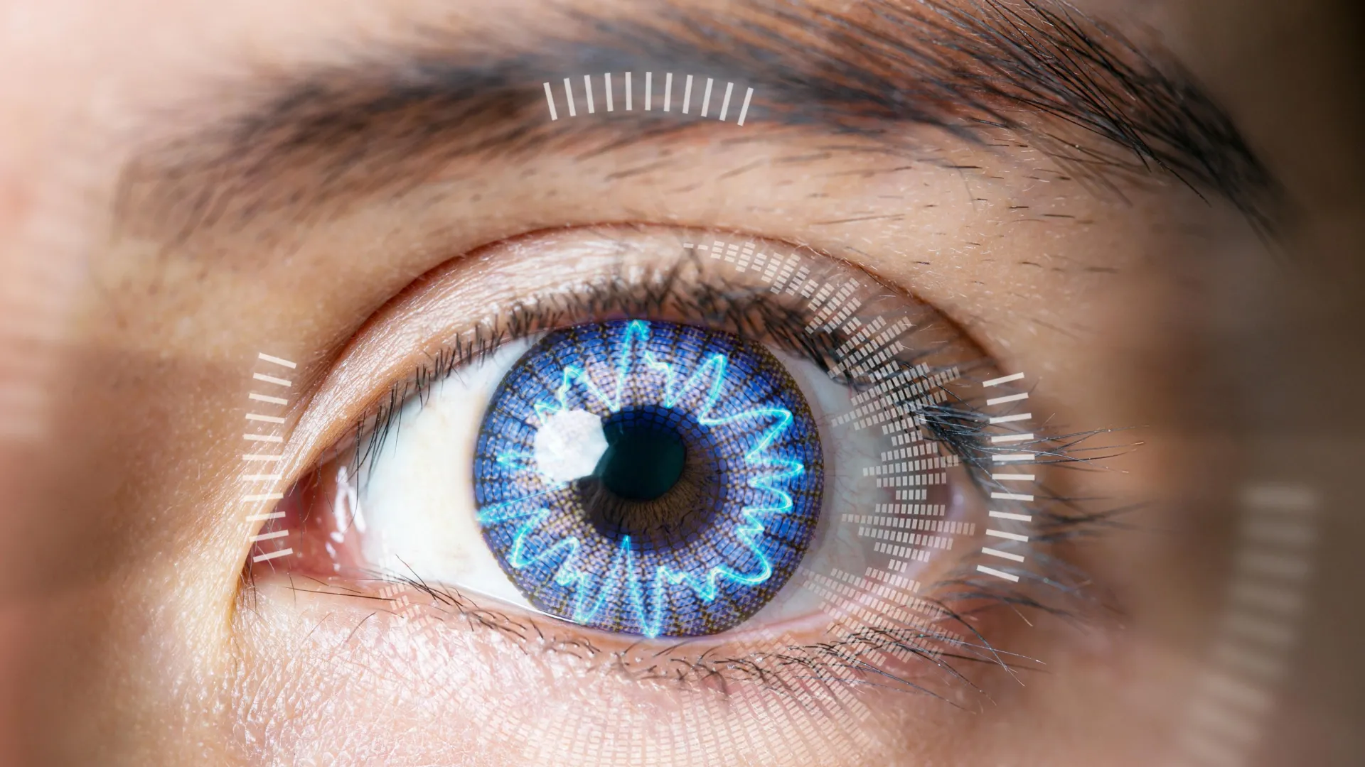
To understand the magnitude of this innovation, one must first appreciate the delicate physics of the human eye. The cornea is the clear, dome-shaped surface that acts as the eye’s outermost lens. It is the primary gatekeeper of vision, responsible for bending (refracting) incoming light so that it converges precisely onto the retina.
The retina, a layer of light-sensitive tissue at the back of the eye, acts as a biological film, converting light signals into neural impulses that the brain interprets as images. When the cornea’s curvature is slightly "off"—too steep, too flat, or irregular—light fails to focus on the retina, resulting in the blurriness characteristic of refractive errors.
Traditional surgery like LASIK corrects these errors by permanently removing layers of corneal tissue to alter its curvature. As Michael Hill, a professor of chemistry at Occidental College, notes, "LASIK is just a fancy way of doing traditional surgery. It’s still carving tissue—it’s just carving with a laser." While generally considered safe, the permanence of tissue removal and the risks associated with surgical incisions have long motivated scientists to seek a more elegant solution.
The Chronology of an Accidental Discovery
The journey toward EMR did not begin in an ophthalmology suite, but rather in a laboratory setting focused on the fundamental properties of living tissues. Brian Wong, a professor and surgeon at the University of California, Irvine, was initially investigating the biomechanics of cartilage when he stumbled upon a surprising phenomenon.
"The whole effect was discovered by accident," Wong explains. "I was looking at living tissues as moldable materials and discovered this whole process of chemical modification."
Wong and his collaborator, Michael Hill, realized that the structural proteins found in cartilage—collagen—were also the primary components of the cornea. Collagen is held together by a complex web of electrically charged components. By applying a controlled electric potential to the tissue, the researchers discovered they could manipulate the pH environment within the collagen matrix.
By lowering the pH, the tissue becomes temporarily acidic, weakening the electrical attractions that keep the collagen fibers rigid. In this "plastic" state, the tissue becomes malleable. Once the pH is returned to a neutral level, the collagen fibers "reset" their bonds, locking the tissue into its new, reshaped form. This discovery marked the transition from theoretical chemistry to a potential clinical breakthrough.
Supporting Data: From Rabbit Ears to Corneal Molds
The research team began their validation process by testing EMR on cartilage-rich rabbit ears, successfully demonstrating that the tissue could be reshaped without trauma. Encouraged by these results, they moved to the eye.
The team engineered specialized platinum "contact lenses" that serve as both a mold and an electrode. In experiments conducted on 12 rabbit eyeballs, these lenses were placed over the corneas while the eyes were submerged in a saline solution mimicking human tears.
The results were striking. Within approximately 60 seconds—a timeframe comparable to the laser portion of a LASIK procedure—the cornea shifted to mirror the exact curvature of the platinum mold. Most significantly, histological analysis revealed that the corneal cells remained healthy and viable post-procedure. The researchers had successfully navigated the "goldilocks zone": the pH change was sufficient to alter the shape of the collagen, but not so extreme as to induce cellular necrosis.
In a subset of 10 "myopic" eyes, the EMR technique achieved the precise optical change required to correct vision. Beyond simple refractive correction, the researchers observed that EMR might be capable of reversing corneal cloudiness, a debilitating condition that currently necessitates risky corneal transplants.
Official Responses and Scientific Peer Review
The scientific community has viewed the research with a mix of cautious optimism and intense interest. While the data remains preliminary, the mechanical simplicity of the EMR system is a major point of appeal. Unlike LASIK, which requires multimillion-dollar laser suites, clean rooms, and extensive post-operative monitoring for healing incisions, EMR is theoretically portable and minimally invasive.
However, the team—funded by the National Eye Institute of the National Institutes of Health and the John Stauffer Charitable Trust—is the first to acknowledge the "long road" ahead. Dr. Wong describes the next phase of the project as a "long march through animal studies that are detailed and precise."
Before the technique can ever reach human trials, the researchers must prove that the reshaping is stable. The eye is a dynamic organ subject to constant pressure and chemical fluctuations; ensuring that the cornea does not "revert" to its original shape over time is the next hurdle. Furthermore, the researchers must investigate whether the procedure can address the full spectrum of human vision issues, including astigmatism and higher-order aberrations.
Implications: A Paradigm Shift in Ophthalmology
If EMR successfully makes the transition from the laboratory to the clinic, it could represent a tectonic shift in how we approach vision health. The implications are three-fold:
1. Democratization of Vision Care
The high cost of LASIK limits the procedure to those with significant disposable income or robust insurance coverage. Because EMR requires significantly less complex equipment and no sterile surgical theater for incisions, the cost of the procedure could be drastically lower, potentially making vision correction accessible to underserved populations globally.
2. Enhanced Safety and Reversibility
Because EMR does not involve the permanent removal of tissue, it introduces the possibility of reversibility. If a patient’s prescription changes or if they are dissatisfied with the results, the cornea could theoretically be reshaped again. This "adjustable" nature of the procedure removes the anxiety associated with the permanence of laser surgery.
3. Broadening the Scope of Treatment
The discovery that EMR can potentially treat corneal cloudiness opens doors to non-surgical treatments for pathologies that currently have few options besides invasive transplantation. If the electrical modulation of collagen can be mastered, it could become a platform technology for treating a variety of structural eye diseases.
The Road Ahead
Despite the potential, the researchers remain grounded. "There’s a long road between what we’ve done and the clinic," Michael Hill admits. "But, if we get there, this technique is widely applicable, vastly cheaper, and potentially even reversible."
For now, the project faces the typical challenges of early-stage biomedical research: funding cycles, the necessity of long-term longitudinal studies in living models, and the stringent regulatory hurdles of the FDA. The transition from "isolated rabbit eyeballs" to a living, blinking human eye is a massive scientific leap.
As the researchers prepare for their next series of animal studies, the medical community watches with bated breath. The age of the laser may be far from over, but for those who dream of a future where vision is corrected with the simple flick of a switch, the "electrifying" potential of EMR offers a glimpse of a clearer tomorrow. The quest to perfect human sight continues, and for the first time in a long time, the solution might not be to cut, but to gently reshape.