
The Silent Crisis of the Stroke Cavity
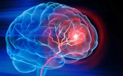
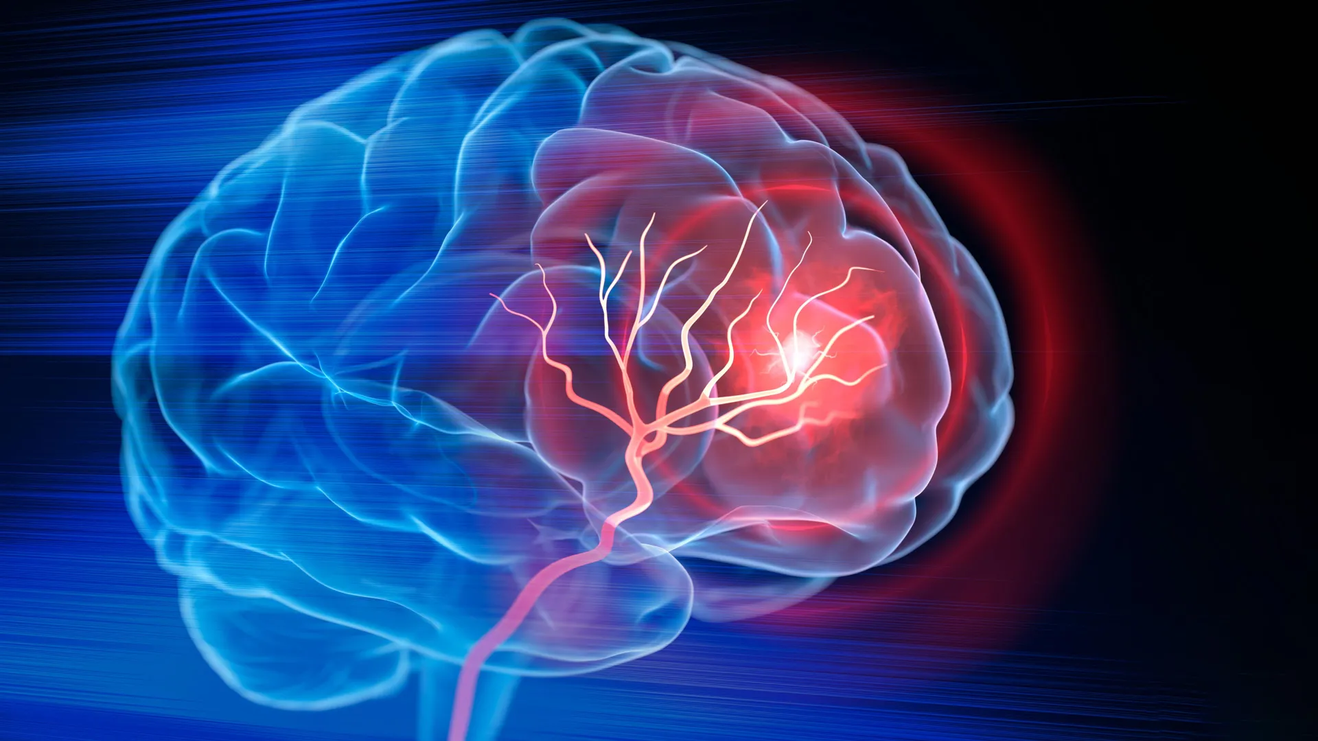
For millions of individuals worldwide, an ischemic stroke marks the beginning of a long, often incomplete journey toward recovery. When a blood clot obstructs the flow of oxygenated blood to the brain, the resulting hypoxia triggers a cascade of cell death. While modern emergency medicine—including mechanical thrombectomies and tissue plasminogen activator (tPA) therapies—has revolutionized the ability to clear blockages and salvage at-risk brain tissue, these interventions are inherently limited by a temporal window. Once the tissue has progressed to necrosis, it leaves behind a permanent, fluid-filled cavity, or "lesion," that effectively severs the neural pathways that once governed movement, cognition, and sensory perception.
Standard post-stroke care has historically focused on rehabilitation—physical, occupational, and speech therapies designed to "retrain" surviving brain circuits to compensate for the lost regions. While effective to a degree, this approach does not address the void left by the necrotic tissue. Now, a pioneering study published in the journal Cell Biomaterials by a team of biomedical engineers at Duke University suggests a paradigm shift: rather than simply working around the damage, we might soon be able to engineer the environment to rebuild it.
A Novel Biomaterial Scaffold: The MAPS Approach
The research, led by Tatiana Segura, the Robert Plonsey Distinguished Professor of Biomedical Engineering at Duke, introduces an injectable biomaterial designed to transform the hostile environment of a stroke cavity into a fertile ground for biological repair.
The core of this technology is a platform known as MAPS, or microporous annealed particle scaffolds. Unlike traditional monolithic hydrogels, which can be dense and impenetrable to cellular infiltration, MAPS are composed of individual, microscopic hydrogel spheres. When injected, these spheres self-assemble into a porous, interconnected structure. This "micro-porosity" acts as a sophisticated scaffold, creating a physical framework that invites the body’s own cells to migrate into the injured space.
"Once brain tissue has been lost, restoring blood flow is no longer enough," Dr. Segura explained. "Our goal is to engineer the injured space so that immune, vascular, and neural repair processes can begin to work together."
Chronology of the Repair Strategy
The research team’s strategy unfolded through a meticulous, multi-stage experimental process:
- The Design Phase: Researchers sought to harness the communicative power of astrocytes—the star-shaped glial cells that serve as the brain’s "first responders." They specifically targeted extracellular vesicles (EVs), which act as biological courier packages containing proteins, lipids, and genetic material.
- The Engineering Phase: Recognizing that injecting therapeutic signals directly into the brain would lead to their rapid diffusion and degradation, the team chemically tethered these signaling molecules to the surface of the MAPS particles. This ensured that the therapeutic cargo remained concentrated within the scaffold, maximizing the opportunity for cellular interaction.
- The Recruitment Phase: The team tested various combinations of signaling molecules to determine which would most effectively trigger healing. They identified a potent combination—interleukin-4 (IL-4) and complement component C1q—that successfully recruited specific immune cell populations into the injury site.
- The Biological Evaluation: Over an eight-week period, the team monitored the structural and functional changes in mouse models, utilizing grid-walking tests to measure motor coordination and recovery.
The Surprising Role of Neutrophils
One of the most significant findings in the study challenges long-held dogmas in neuroimmunology regarding neutrophils. Historically, these white blood cells have been viewed primarily as "villains" in the context of stroke, contributing to acute inflammation and secondary tissue damage in the immediate aftermath of an ischemic event.
However, the Duke team’s findings suggest that the role of the neutrophil is highly context-dependent. By providing a stable, signaling-rich environment within the MAPS scaffold, the researchers observed that neutrophils could be redirected to serve a constructive purpose. When the team experimentally depleted the neutrophil population in the treated mice, they witnessed a marked decline in both blood vessel formation and scaffold remodeling.
"This result changes how we think about neutrophils after stroke," said Shangjing Xin, the study’s lead scientist and a postdoctoral fellow in the Segura Laboratory. "Their role appears to depend on when they arrive, where they are located, and the signals they receive from their surroundings. Our study demonstrates a potential engineering strategy to recruit and retain these cells at the right time."
Data-Driven Results: From Molecular Signaling to Motor Function
The data gathered from the study provides a compelling case for the efficacy of the MAPS-EV system. The results were categorized into three key metrics of success:
- Vascularization: The scaffold-injected animals showed a significant increase in the formation of new blood vessels, essentially "re-vascularizing" the stroke cavity. This is a critical step, as oxygen delivery is mandatory for the survival and growth of new neural tissue.
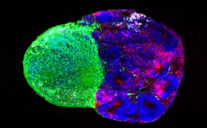
- Neural Connectivity: Immunofluorescent imaging revealed a higher density of axonal fibers—the "cables" that allow neurons to communicate—both within the scaffold and in the peri-infarct region (the border zone surrounding the injury).
- Functional Restoration: The most striking evidence of recovery came from the grid-walking test. Mice treated with the optimized MAPS scaffold showed a marked reduction in forelimb placement errors. By the eight-week mark, their performance was statistically indistinguishable from healthy control subjects, a level of functional recovery that persisted for the duration of the study.
Crucially, the team conducted control experiments to see if the extracellular vesicles alone, without the scaffold, could achieve these results. They could not. The physical architecture of the MAPS scaffold was found to be essential, as it provided the necessary structural substrate for cell adhesion and ensured the prolonged, localized release of therapeutic signals.
Implications for Future Clinical Applications
While the results are undeniably promising, the researchers are quick to emphasize that this remains a preclinical breakthrough. The current study was performed in mice, and translating this technology to human clinical settings presents several hurdles.
Safety and Scaling
One primary challenge is the source of the extracellular vesicles. The current study utilizes primary rat astrocytes, which is not a sustainable or viable path for human clinical trials. The team is currently pivoting to human induced pluripotent stem cell (iPSC)-derived astrocytes. By generating these cells in a laboratory setting, the researchers hope to gain greater control over the composition of the EVs while ensuring a scalable source for future therapies.
Human-Scale Modeling
Furthermore, the anatomy and physiology of a human brain are vastly more complex than that of a mouse. Larger animal models will be required to determine if the physical volume of a human stroke cavity can be effectively bridged by this hydrogel technology and to assess the long-term safety of introducing such materials into the human central nervous system.
A New Philosophy of Neuroregeneration
Dr. Segura’s vision for the future of stroke treatment is as much a philosophical shift as it is a technological one. She compares the process to ecological restoration.
"You do not restore an ecosystem simply by containing the initial damage," Segura stated. "You have to create the conditions that allow life to return. That is how we think about the stroke cavity. The material is not intended to reproduce the brain itself, but to create an environment where the body’s own cells can enter, communicate and participate in rebuilding vascularized tissue."
By viewing the stroke cavity not as a dead end, but as a site for potential reconstruction, the Duke team has opened a new door in regenerative medicine. If the findings can be successfully translated to human trials, this "environmental engineering" approach could move medicine away from merely mitigating the impact of a stroke and toward actively repairing the damage, potentially offering a path to recovery for millions who were previously told their brain injury was permanent.
The path ahead involves rigorous safety testing and refining the signaling "cocktails" delivered via the scaffolds, but the implications are clear: the future of stroke recovery may lie in our ability to work with the body’s own biological machinery, providing it with the right structure and the right instructions to heal itself from within.