
For decades, the human brain was viewed as a static, insulated organ, protected by the rigid blood-brain barrier and shrouded in biological mystery. However, since the 2012 discovery of the "glymphatic system" by pioneering neuroscientist Maiken Nedergaard of the University of Rochester, our understanding of neurological health has undergone a seismic shift. We now know that during deep, restorative sleep, the brain undergoes a rigorous "wash cycle." A water-like fluid—cerebrospinal fluid (CSF)—surges through the brain’s architecture, flushing out metabolic waste, including the sticky amyloid-beta proteins implicated in the progression of Alzheimer’s disease.
Despite the monumental nature of this discovery, the mechanics of this system have remained frustratingly elusive. Scientists have long struggled to quantify exactly how this fluid moves through the dense, gelatinous landscape of the brain. Now, a breakthrough study published in Science Advances by an interdisciplinary team from the University of Rochester, Brown University, and the University of Copenhagen has bridged this gap. By deploying physics-informed artificial intelligence, researchers have successfully mapped the velocity of brain fluid, revealing a complex, two-speed system that could one day revolutionize how we diagnose and treat neurodegenerative disorders.
The Chronic Challenge: Visualizing the Invisible
The primary hurdle in glymphatic research has always been the "scale paradox." On one hand, high-resolution optical microscopy allows researchers to observe a tiny patch of brain tissue with exquisite clarity. However, this method provides only a "keyhole" view—a microscopic observation that fails to account for the systemic movement of fluid across the entire brain.
Conversely, clinical tools like Magnetic Resonance Imaging (MRI) provide a comprehensive, three-dimensional view of the living brain, making them the gold standard for diagnostic imaging. Yet, traditional MRIs are notoriously poor at capturing the movement of fluids at the microscopic speeds inherent to the glymphatic system. In the brain, fluid does not flow like a river; it creeps like a slow-moving tide through a porous sponge. Measuring these "glacial" velocities without causing surgical damage to the subject has been the holy grail of neuro-engineering.
"You can put a microscope on a small patch of the brain and watch what’s happening there with a lot of detail, and we’ve worked with that type of data in the past, but it’s only a tiny view of the overall process," explains Professor Douglas Kelley, a fluid dynamics expert from the University of Rochester’s Department of Mechanical Engineering. "If you want to image whole brains, an MRI is a great approach because it gives you a three-dimensional view. But an MRI has serious limitations too, the biggest of which is that it does not capture the fluid flow velocity, at least not for flows this slow."
Chronology of a Breakthrough: From Biological Mystery to AI Mapping
The path to this discovery was paved by a multi-year effort to integrate fluid mechanics with machine learning. The researchers sought to overcome the limitations of MRI by creating a "physics-informed" neural network—a form of AI that doesn’t just guess patterns, but respects the fundamental laws of physics that govern how fluids move through porous materials.
- Phase I: Data Collection: The team began by analyzing videos of dye diffusing through brain tissue. These videos provided the "ground truth"—a visual representation of how particles travel through the tissue’s complex geometry over time.
- Phase II: Neural Network Training: Using this data, the team trained AI models to recognize the relationship between dye dispersal and fluid speed. By feeding the model the physics of fluid dynamics, the AI learned to predict how quickly fluid must be moving to push the dye in the observed patterns.
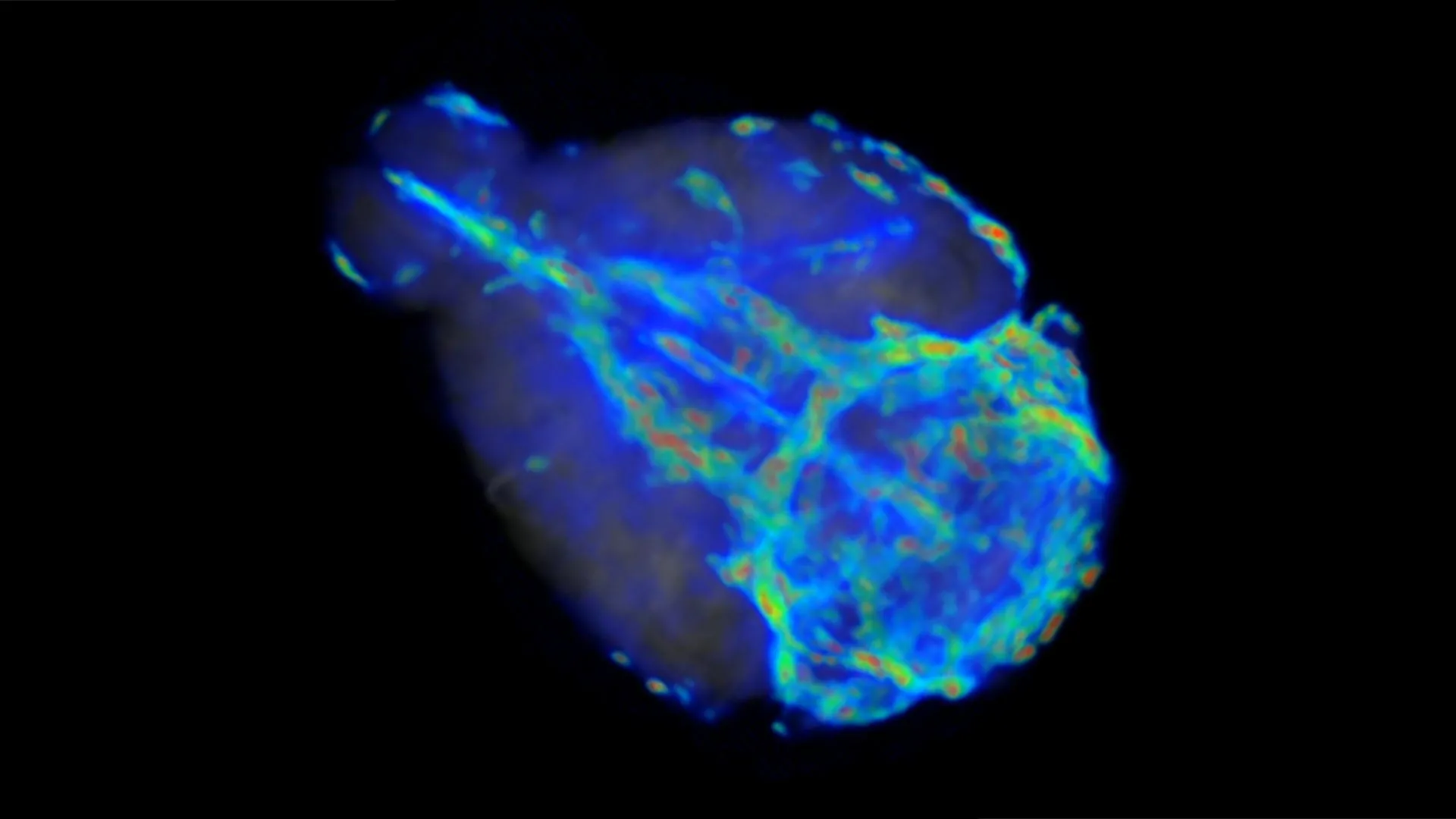
- Phase III: Integration with MRI: With the model trained, the team applied it to MRI data. The AI acted as a digital lens, "de-blurring" the MRI images to reveal the underlying flow velocities, effectively calculating both the speed of the fluid and the permeability of the surrounding tissue.
This chronological progression from raw observational data to sophisticated, physics-informed computation marks a new era in neuro-imaging, where the limitations of hardware are bypassed by the capabilities of software.
Two Speeds of the Mind: Supporting Data
The findings of the study were striking. The team discovered that the glymphatic system is not a monolithic flow, but rather a dual-speed network.
In the more open, interstitial spaces—particularly the areas near the surface of the brain, between the skull and the brain matter—the cerebrospinal fluid flows at a relatively brisk pace of a few microns per second. While "brisk" is a relative term at the microscopic level, it is significantly faster than the fluid movement found deeper within the brain.
Deep inside the dense parenchyma—the functional tissue of the brain—the fluid encounters massive resistance. Here, the flow is roughly 50 times slower than the surface circulation. This discovery explains why the brain is so vulnerable to the accumulation of toxic proteins: the deeper the region, the more difficult it is for the "wash cycle" to reach it and clear away metabolic debris. These data points provide the first quantitative baseline for understanding how efficiently the brain cleans itself during sleep, offering a clear metric against which diseased states can be compared.
Official Responses and Collaborative Effort
The research was a massive collaborative undertaking, supported by the NIH National Center for Complementary and Integrative Health and the NIH BRAIN Initiative. This level of support underscores the high stakes of the project: the potential to manage, or even prevent, the global Alzheimer’s epidemic.
The research team, which includes a diverse group of computational scientists, neurobiologists, and engineers, emphasizes that this is only the beginning. The list of contributors highlights the interdisciplinary nature of the work: Brown University PhD student Juan Diego Toscano, URochester computational scientist Yisen Guo, Brown University PhD student Zhibo Wang, URochester PhD student Mohammad Vaezi, University of Copenhagen Associate Professor Yuki Mori, Brown University Professor George Karniadakis, and URochester Assistant Professor Kimberly Boster.
Their collaborative response has been one of cautious optimism. While the physics-informed AI is a robust tool for mice, the team is already looking toward the horizon of human application. By establishing these baseline measurements in animal models, they are refining the AI to be sensitive enough to detect the subtle differences between a healthy brain and one beginning to show signs of cognitive decline.
Clinical Implications: The Future of Neurological Care
The long-term goal of this research is to bring this technology into the clinic. If doctors can measure the "hydraulic health" of a patient’s brain, it could change the landscape of neurology as significantly as the invention of the blood pressure cuff changed cardiology.
Early Detection and Screening
The most exciting application is early screening. Currently, Alzheimer’s is often diagnosed only after significant cognitive impairment has occurred. If physicians could use an MRI-based AI scan to detect "sluggish" glymphatic flow in a 40- or 50-year-old, it might provide a decades-long window for intervention—whether through lifestyle changes, sleep hygiene optimization, or pharmacological treatments designed to boost fluid circulation.
Assessing Brain Trauma
Beyond neurodegeneration, the study has profound implications for traumatic brain injury (TBI). Concussions and other head injuries are known to disrupt the brain’s internal environment. "We could check when somebody has been concussed to see whether the fluid circulation in their brain is disrupted," says Professor Kelley. Being able to track the recovery of fluid flow could help clinicians determine when a patient is truly healed, rather than relying on subjective reports of symptom resolution.
A New Diagnostic Paradigm
The integration of physics-informed AI with standard clinical imaging means that this technology could be deployed without requiring new, invasive equipment. By simply updating the software on existing MRI machines, hospitals could eventually gain the ability to visualize the brain’s waste-removal system in real-time.
As the researchers continue to refine their models, they are moving closer to a future where "brain washing" is not just a biological process, but a clinical metric. By understanding the speed, rhythm, and flow of our brain’s midnight cleanup, we are finally moving toward a world where we can protect the mind from the silent, creeping buildup of the diseases that threaten our golden years. This study serves as a critical bridge between the theoretical discovery of the glymphatic system and the practical, life-saving diagnostic tools of tomorrow.