
Congenital heart disease (CHD) stands as the most prevalent birth defect globally, impacting approximately one in every 100 newborns. For decades, the medical community has grappled with the mystery of why certain genetic mutations result in such devastating structural anomalies. While researchers have long identified specific genes—such as TBX5—as critical architects of the heart, the "why" behind the clinical severity remained elusive. A groundbreaking study published in the journal Science by researchers at the Gladstone Institutes has finally pulled back the curtain, revealing that the problem isn’t just the code itself, but the way that code is physically folded within the cell.
The Mystery of Haploinsufficiency
At the heart of this research is a phenomenon known as "haploinsufficiency." In a healthy individual, two copies of every gene are inherited—one from each parent. In many cases of congenital heart disease, a child is born with only one functional copy of a gene, such as TBX5.
For years, the biological question has been: why does the loss of just one copy have such a profound, clinical impact when a second, healthy copy remains active? Traditional genetic dogma suggested that the single remaining copy might simply be unable to produce enough of the necessary protein to sustain normal development. However, the Gladstone team suspected a more structural, mechanical failure. They hypothesized that the protein encoded by TBX5 does more than just switch genes on and off; it acts as an essential "structural engineer" for the genome itself.
The 3D Instruction Manual: How DNA Folding Works
To understand the significance of this discovery, one must first grasp the complexity of DNA storage. If stretched out, the DNA in a single human cell would be nearly two meters long. To fit this massive volume of information into the microscopic space of a cell nucleus, the body uses a sophisticated, hierarchical folding system.
This process is not random. The cell arranges its genetic material into a highly specific three-dimensional architecture, akin to a meticulously organized instruction manual. This organization occurs in layers:
- Compartments: The largest units, functioning like separate binders of information.
- Domains: Sub-units akin to individual paragraphs of instructions.
- Chromatin Loops: The most intricate level, where distant segments of DNA are brought together to allow "enhancers"—genetic switches—to physically touch and activate specific genes.
This 3D structure is the primary reason why a heart cell functions differently than a brain cell; even though they share the same DNA, they access different "pages" of the manual because their 3D configuration keeps certain instructions open and others tucked away.
Chronology: Unraveling the Genetic Blueprint
The journey to this discovery began by synthesizing stem cells into functional heart muscle cells. By creating three distinct groups—healthy cells, cells lacking one copy of TBX5, and cells lacking both copies—the researchers were able to observe the "before, during, and after" of heart cell development.
Phase 1: High-Resolution Mapping
The team utilized advanced, high-resolution 3D mapping techniques to visualize the chromatin loops at an unprecedented level of detail. Because the process generated millions of data points, the team turned to cutting-edge computational modeling. "We developed and used different computational models to analyze results from thousands of individual cells," explains Dr. Katie Pollard, director of the Gladstone Institute of Data Science and Biotechnology. "That allowed us to finally see how losing this one protein causes the heart’s DNA structure to break down on every level."
Phase 2: Identifying the Molecular Architect
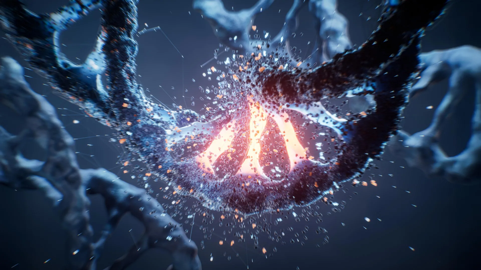
As the stem cells matured into heart muscle, the researchers observed the genome undergoing major structural shifts. They identified TBX5 as the central organizer of these changes. Specifically, the protein acts as a GPS system for "cohesin," a molecular motor that moves along the DNA to pull it into the necessary loops. When TBX5 levels drop, the cohesin motor loses its guidance, the loops fail to form, and the 3D architecture collapses.
Phase 3: The Dose-Dependent Collapse
Perhaps the most striking revelation of the study was the sensitivity of the system to the concentration of the TBX5 protein. The team found that the damage was dose-dependent: the more TBX5 was removed, the more catastrophic the disruption to the genome’s folding. Reducing the protein to half its normal amount—the exact state of a patient with haploinsufficiency—was sufficient to trigger a breakdown across all levels of organization.
Supporting Data and Technical Insights
The research relied on an immense dataset, allowing for a granularity never before achieved in cardiovascular genetics. By analyzing individual cells, the team discovered that not all cells respond uniformly to the loss of TBX5.
"Surprisingly, we discovered this collapse happens at every level of genome organization—compartments, domains, and chromatin loops," says Dr. Shuzhen Kuang, a first author of the study.
Furthermore, the data showed significant heterogeneity between different types of heart cells, such as atrial versus ventricular cells. This variation provides a potential answer to a long-standing clinical enigma: why two individuals with the exact same genetic mutation might exhibit widely different heart defects. The "3D folding error" appears to be influenced by the specific cellular environment, offering a new frontier for personalized medicine and developmental biology.
Official Responses and Expert Perspectives
The study’s senior authors, Dr. Benoit Bruneau and Dr. Katie Pollard, believe this work marks a paradigm shift in how we categorize developmental disorders.
"TBX5 is just one example of a broader class of genes that cause birth defects when only one copy is lost," says Dr. Bruneau, director of the Gladstone Institute of Cardiovascular Disease. "What’s exciting about our findings is they suggest many different birth defects might happen for the same reason: the cell’s 3D instruction manual simply gets folded the wrong way."
Dr. Zoe Grant, a postdoctoral researcher in the Bruneau lab and a first author of the study, emphasized the mechanical nature of the discovery: "What was striking was how the amount of TBX5 matters immensely. The more TBX5 you removed, the worse the disruption across every level of genome organization we looked at."
Clinical and Scientific Implications
The implications of this study reach far beyond congenital heart disease. By identifying a new mechanism of disease—structural misfolding of the genome due to protein scarcity—the researchers have opened the door to re-evaluating numerous other developmental disorders.
1. A New Framework for Genetic Diagnosis
Current genetic testing often focuses on identifying mutations in protein-coding sequences. This research suggests that clinicians may eventually need to look for "folding mutations"—changes that do not necessarily break a protein, but break the coordination required to organize the genome.
2. Potential for Future Therapeutics
If a disease is caused by a failure of 3D folding, the potential for intervention changes. Rather than just focusing on gene therapy to replace a missing protein, researchers might eventually look for ways to stabilize the chromatin structure or recruit molecular motors like cohesin to the correct locations, effectively "re-folding" the genome to a functional state.
3. Future Research Directions
The team at Gladstone is already looking ahead. Their next phase of study aims to determine the precise timeline of when TBX5 begins its organizational duties during embryonic development. They also intend to investigate whether other proteins associated with birth defects perform similar "architectural" functions, which could suggest a universal, previously unrecognized system of genome regulation.
As the scientific community digests these findings, one thing is clear: the instruction manual for human life is not just about the text written on the pages, but about how those pages are bound and organized. For the millions affected by birth defects, this new understanding of the "3D genome" offers a flicker of hope that we may one day learn how to fix the book when the binding fails.
Study Reference:
Grant, Z. L., Kuang, S., et al. (2026). "Dose-dependent sensitivity of human three-dimensional chromatin to a heart disease-linked transcription factor." Science.
Acknowledgments:
This work was supported by the National Institutes of Health (NHLBI U01 HL157989, UM1HG011585, R01 HL155906), the California Institute for Regenerative Medicine, Additional Venture, the Roddenberry Foundation, the Younger Family Fund, and the Saving tiny Hearts Society.