
In the microscopic landscape of the human body, epithelial tissues serve as the ultimate sentinels. Forming the continuous, sealed layers that line our skin, gut, and airways, these cells act as a vital barrier against the outside world. For decades, biologists have understood the primary role of the protein E-cadherin: it acts as the "molecular glue" that tethers these cells together, providing the structural integrity required to keep our tissues intact.
However, a groundbreaking study published in Nature Communications has unveiled an unexpected, dual identity for this essential protein. Researchers have discovered that the E-cadherin complex does not merely maintain the barrier; it orchestrates the systematic engulfment and removal of dead cells from within the tissue. This revelation shifts our fundamental understanding of tissue homeostasis and offers a provocative new lens through which to view chronic inflammation and immune defense.
The Discovery: A Shift in Cellular Responsibility
The research, led by ICREA Research Professor Verena Ruprecht, sought to understand how tissues maintain their integrity while simultaneously managing the inevitable "cellular waste" generated by apoptosis, or programmed cell death. When a cell dies within a tissue, it must be removed efficiently to prevent the leakage of intracellular contents, which can trigger harmful inflammatory responses.
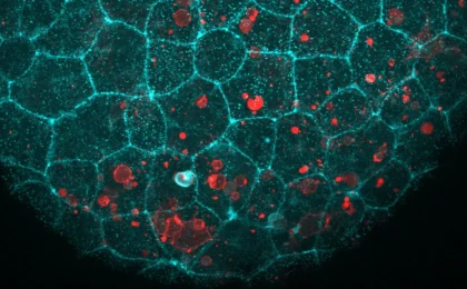
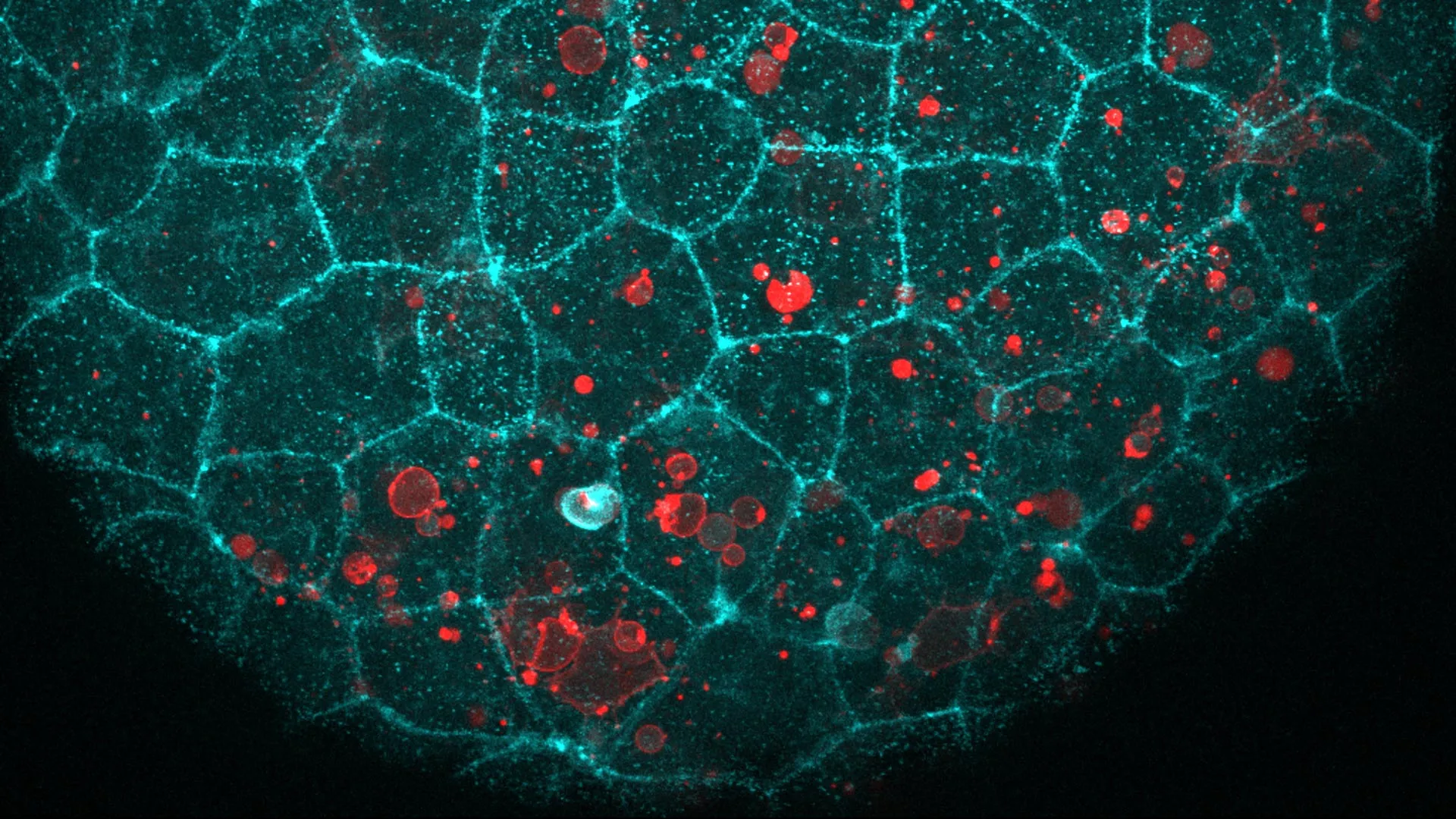
Using high-resolution live imaging in zebrafish and mouse embryos, the team observed a curious phenomenon. When a cell reached the end of its life cycle, the very machinery responsible for cell-to-cell adhesion—the E-cadherin complex—migrated to the precise site of the dying cell.
"We were intrigued to find out that epithelial cells repurpose their molecular adhesion machinery—the ‘glue’ that normally holds them together—to engulf dying cells," says Ruprecht. This finding suggests that the body is remarkably efficient, utilizing pre-existing infrastructure to perform high-stakes maintenance rather than deploying specialized immune cells for every minor cleanup task.
Chronology of the Investigation
The path to this discovery was paved by a series of rigorous experiments designed to test whether E-cadherin was truly the "driver" of this process or merely a bystander.
Phase 1: Identifying the Mechanical Link
The researchers first examined whether the E-cadherin complex relied on specific molecular recognition to identify dead cells. To test this, they introduced "stripped" dying cells—cells from which E-cadherin had been removed—into the epithelial tissue. Surprisingly, the tissue cleared these cells just as effectively as normal, intact dying cells.
Phase 2: Testing the "Eat-Me" Signal
To further refine their understanding, the team introduced artificial fat droplets into the tissue. These droplets lacked the complex protein structures of a real cell but were coated with specific signals normally displayed on the surface of dying cells. The epithelial cells treated these droplets as debris, engulfing them with the same efficiency as biological waste. This confirmed that the E-cadherin complex is not "reading" the identity of the cell, but rather responding to a broader mechanical and chemical "eat-me" signal.
Phase 3: The Mechanical "Dancer"
Perhaps the most significant breakthrough was observing how a cell manages to "eat" its neighbor without compromising the integrity of the tissue barrier. If an epithelial cell were to pull away from its neighbors to swallow a dead cell, the entire barrier could theoretically breach.
Through live imaging, the researchers uncovered a sophisticated, asymmetric response. The upper surface of the epithelial cell—the side exposed to the lumen or the outside environment—remained remarkably stable and rigid. Meanwhile, the lower surface, hidden within the tissue, underwent dramatic deformation, stretching and bending to envelop the debris.
Ruprecht compares this to a row of dancers holding hands: "Their upper bodies remain steady while their feet perform increasingly complicated movements when a dying cell appears. It’s the same dancer with a different choreography."
Supporting Data: The Mechanics of Molecular Ropes and Brakes
To understand how this choreography is physically performed, the research team analyzed the components of the E-cadherin complex, identifying two critical elements that govern the "cleanup" process.
The Rope: Linking to the Skeleton
One specific protein within the complex acts as a tether, or "rope," connecting the E-cadherin assembly to the cell’s internal skeleton (the cytoskeleton). This linkage is vital; it allows the cell to transmit the force required to pull the debris into its interior. When the researchers genetically removed this tethering protein, the cells lost the ability to pull in the dead material, resulting in the accumulation of cellular debris.
The Brake: Regulating Contraction
The second component acts as a "brake" on the cell’s contractile machinery. In a counterintuitive twist, researchers found that removing this brake did not accelerate the cleanup process. Instead, it made the epithelial cells too stiff. Without the ability to regulate their own tension, the cells were unable to deform properly to accommodate the dead cell, proving that the cleanup process requires a delicate balance of tension and flexibility.
The Evolutionary Perspective: A Shared Vertebrate Mechanism
The significance of these findings is bolstered by their consistency across species. By investigating early mouse embryos, the team determined that when E-cadherin function was blocked, dead cells remained trapped in the tissue. This result, mirroring the observations in zebrafish, suggests that this mechanism is not a quirk of one organism, but a deeply conserved, shared trait among vertebrates.
This builds upon previous work by Ruprecht, which identified that embryonic epithelial tissues act as a form of "early innate immune defense." Before a complex immune system fully develops in an embryo, the epithelial cells themselves take on the burden of keeping the tissue clean, preventing the potential toxicity of decaying cellular material.
Implications for Human Health and Chronic Inflammation
While the study focused on embryonic models, the implications for adult human health are profound. Chronic inflammation is a hallmark of many modern ailments, ranging from autoimmune disorders to neurodegenerative diseases.
Understanding Failure
If the cleanup mechanism—the "garbage disposal" system of the epithelium—fails, dead cells are left to linger and eventually rupture. This "secondary necrosis" releases cellular components into the surrounding tissue, which the immune system recognizes as a sign of danger, triggering a cascade of inflammation. By understanding how the E-cadherin complex functions in a healthy state, researchers may eventually identify why these processes fail in diseased states.
Broadening the Scope
While it remains to be proven if this specific E-cadherin-dependent mechanism is active in adult human tissues, the indicators are promising. Epithelial cells in the retina, colon, and airways are already known to clear debris. Furthermore, the structural consistency of E-cadherin across the animal kingdom suggests that this "cleanup crew" may be a fundamental, ancient feature of all epithelial tissues.
"Studying the mechanisms of how dying cells can be removed efficiently from tissues is of very high relevance to human health," says Ruprecht. "We are essentially looking at the maintenance crew of the human body."
Conclusion: A New Era for Tissue Engineering
This study represents a significant leap forward in cell biology, moving from a static view of cells as "bricks" in a wall to a dynamic view of them as active participants in their own environment’s maintenance. By revealing that structural proteins can moonlight as cleanup agents, the research provides a new foundation for future therapeutic interventions.
As the scientific community continues to explore the nuances of this "cellular choreography," the focus will shift toward whether we can modulate this process in adults. If we can enhance or repair the "cleanup" efficiency of tissues, we might find new ways to treat conditions characterized by chronic, unresolved inflammation, ultimately helping the body keep its internal barriers not just sealed, but clean.
Study Credits:
The research was led by joint first authors Hanna-Maria Häkkinen, Marta Batet Palau, and Laura F. Bianchi, under the supervision of Verena Ruprecht. Financial support was provided by the Spanish Ministry of Science and Innovation, the Human Frontier Science Program, the European Union’s Horizon Europe program, and the "la Caixa" Foundation, with additional support from the European Social Fund. Technical support was provided by the CRG Core Facilities for Advanced Light Microscopy, Tissue Engineering, and Protein Technologies.