
For decades, the neuroscientific community has been obsessed with the “hardware” of the brain: the intricate firing of neurons, the architecture of synaptic connections, and the genetic instructions that dictate cellular behavior. Yet, while researchers have mapped the brain’s genes and proteins with pinpoint accuracy, a foundational element of biological life—the humble lipid—has remained largely in the shadows.
These fatty molecules are far more than mere cellular filler. They form the membranes that define the boundaries of every neuron, construct the myelin sheaths that insulate our neural fibers, and act as the essential messengers in cellular communication. Despite their ubiquity, lipids have been notoriously difficult to visualize, leaving a significant “lipid-shaped hole” in our understanding of brain biology.
Now, a pioneering team of researchers at EPFL (École Polytechnique Fédérale de Lausanne) has finally pulled back the curtain. By creating the world’s first high-resolution "Lipid Brain Atlas," scientists have revealed that the brain’s chemical landscape is not a vague, uniform smear, but a highly structured, organized, and dynamic geography that holds the keys to understanding everything from maternal brain adaptation to the progression of Alzheimer’s disease.
The Chronology of a Chemical Breakthrough
The journey to mapping the brain’s lipidome began with a fundamental technological limitation. Conventional imaging techniques, such as MRI or traditional fluorescence microscopy, are adept at capturing anatomical structures or gene expression, but they struggle to differentiate between the subtle variations of individual lipid molecules. For years, this meant that while scientists knew lipids were important, they had little more than a “blurry picture” of their distribution.
The breakthrough, recently published in the journal Nature, was the result of a multi-year effort led by Giovanni D’Angelo, Luca Fusar Bassini, and Gioele La Manno. The process followed a rigorous timeline of innovation:
- Methodological Refinement: The team harnessed the power of mass spectrometry imaging. This technique allows for the precise identification of molecules by scanning thin sections of brain tissue with a laser, mapping the chemical “fingerprint” of every microscopic location.
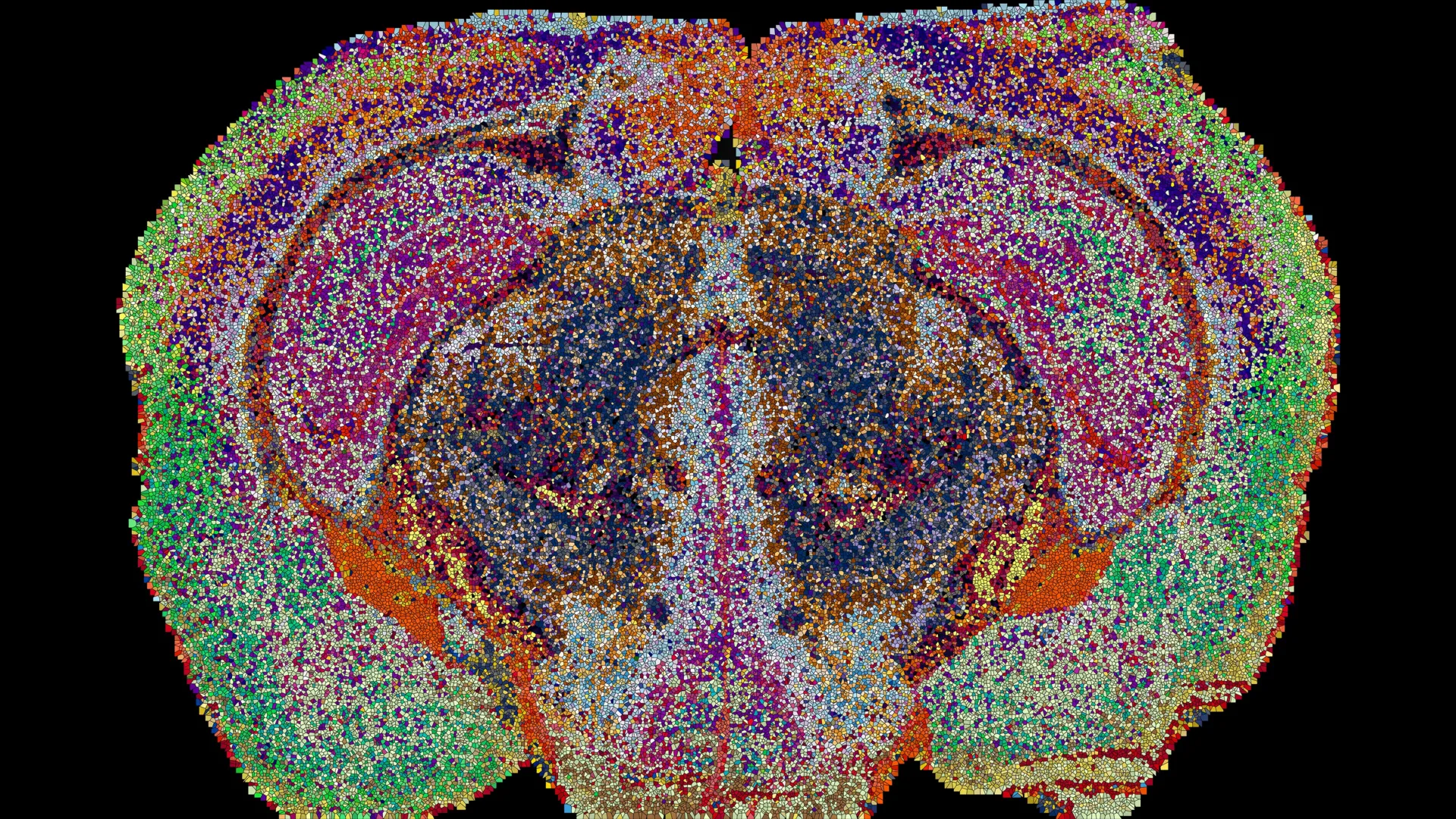
- Data Acquisition: The researchers analyzed 109 brain slices collected from 11 mice. This intensive process yielded approximately 7 million individual data points, creating a dataset of unprecedented scale.
- Algorithmic Synthesis: Recognizing that the raw data was too complex for human interpretation, the team developed custom machine learning tools. These algorithms were designed to stitch together the 2D slices into a comprehensive 3D reconstruction, identifying chemical patterns that were invisible to the naked eye.
- Verification and Mapping: Once the 3D model was complete, the team cross-referenced these chemical territories with known anatomical structures, confirming that their “lipizones” were not merely artifacts of the imaging process but real, functional zones of the brain.
Supporting Data: The Anatomy of ‘Lipizones’
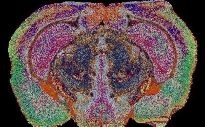
The most striking revelation of the EPFL study is the discovery of 539 chemically distinct territories within the mouse brain, which the researchers have dubbed “lipizones.”
The sheer granularity of these findings challenges the traditional view of brain anatomy. While many of these zones align with known biological structures—such as the hippocampus or the thalamus—others reveal entirely new boundaries. In some instances, the lipid composition is so specific that researchers can identify the origin of a tissue sample simply by analyzing its lipid profile.
The Postal Code Hypothesis
One of the most compelling insights from the data is the "postal code" theory proposed by Giovanni D’Angelo. The atlas suggests that lipids act as chemical addresses. Distant regions of the brain that are functionally related often share the same lipid signature. This suggests that the brain uses its chemical composition to group related cells together, even when they are physically separated by large distances. This form of chemical organization, often invisible to maps focused solely on gene activity, provides a new framework for understanding how the brain synchronizes complex tasks.
Official Responses and Expert Perspectives
The lead researchers view this atlas as a paradigm shift in how we approach neuroscience.
Giovanni D’Angelo, holder of The Kristian Gerhard Jebsen Foundation Chair in Nutrition and Metabolism at EPFL, emphasizes the clinical urgency of this work. "That gap [in our knowledge] matters," he says. "Changes in brain lipids are increasingly linked to conditions from depression to Alzheimer’s disease." By finally establishing what a “normal” lipid map looks like, the researchers have provided a baseline against which pathological changes can be measured.
Luca Fusar Bassini, a key contributor to the study, points to the surprising diversity found within what was previously considered “uniform” tissue. "This shows that the cells which make myelin are far more varied in their chemistry than their genes alone would suggest," he notes. The discovery of distinct lipid zones within white matter—the brain’s “cabling”—suggests that the support cells responsible for insulation are far more specialized than previously assumed.
Gioele La Manno highlights the dynamic nature of these chemical signatures, particularly in the context of physiological changes. "The atlas reveals that the brain constantly adjusts its lipids to meet the body’s changing needs," he explains.
Implications: The Plasticity of the Chemical Brain
Perhaps the most startling findings in the study relate to how the brain’s lipid architecture shifts during pregnancy. By comparing the brains of pregnant mice to those of non-pregnant controls, the team observed widespread, systemic changes in lipid distribution.
These changes were not minor adjustments; they were more profound than the typical differences observed between male and female brain chemistry. Specifically, the cortex—the seat of higher-order cognitive function—underwent extensive chemical remodeling. Simultaneously, the researchers observed a sharp increase in galactosyl ceramide, a critical component of myelin, within the white matter.
These findings suggest that the maternal brain is in a state of constant chemical flux, "retuning its own wiring" to adapt to the physiological demands of pregnancy. This implies that our brain’s "operating system" is not static; it is a flexible, adaptive landscape that leverages lipid metabolism to recalibrate its neural connections.
The Future: From Mice to Medicine
While the current atlas is specific to the mouse brain, the implications for human health are immense. The researchers are quick to clarify that the map itself is not a 1:1 translation for the human brain, but the methodology is a universal key.
A New Diagnostic Frontier
The Lipid Brain Atlas is now freely available online, inviting the global scientific community to integrate this data with existing genetic and cellular maps. For clinicians, this is a game-changer. If researchers can identify how lipid signatures break down in models of Alzheimer’s, Parkinson’s, or depression, they may eventually be able to use lipid profiles as biomarkers for early detection.
Redefining Brain Aging
As the global population ages, understanding the decline of neurological function is a priority. Lipid degradation is a hallmark of many neurodegenerative diseases. With the reference map provided by the EPFL team, scientists can now investigate whether specific "lipizones" are more vulnerable to age-related decline than others.
"What transfers to humans isn’t the map, it’s the method," says Bassini, "and the fact that we now know that a healthy brain has a very organized lipid map at all."
Conclusion: A New Language for Neuroscience
The creation of the Lipid Brain Atlas represents the end of the "blurry" era of lipid research. By demonstrating that the brain possesses a precise, organized, and adaptive chemical geography, the EPFL team has opened a new chapter in biology.
We now have the tools to look beyond the firing of neurons and the expression of genes to examine the fatty scaffolding that makes all of it possible. Whether it is understanding the neural adaptation of pregnancy or unlocking the mysteries of neurodegeneration, the future of brain science is increasingly being written in the language of lipids. The "postal codes" of the brain have finally been mapped, and the discovery is just beginning.